 Movie
Movie Controller
 Structure viewers
 About Yorodumi Papers


+Search query
-Structure paper
| Title | Three-dimensional structure of the native spliceosome by cryo-electron microscopy. |
|---|---|
| Journal, issue, pages | Mol Cell, Vol. 15, Issue 5, Page 833-839, Year 2004 |
| Publish date | Sep 10, 2004 |
 Authors Authors | Maia Azubel / Sharon G Wolf / Joseph Sperling / Ruth Sperling /  |
| PubMed Abstract | Splicing of pre-mRNA occurs in a multicomponent macromolecular machine--the spliceosome. The spliceosome can be assembled in vitro by a stepwise assembly of a number of snRNPs and additional proteins ...Splicing of pre-mRNA occurs in a multicomponent macromolecular machine--the spliceosome. The spliceosome can be assembled in vitro by a stepwise assembly of a number of snRNPs and additional proteins on exogenously added pre-mRNA. In contrast, splicing in vivo occurs in preformed particles where endogenous pre-mRNAs are packaged with all five spliceosomal U snRNPs (penta-snRNP) together with other splicing factors. Here we present a three-dimensional image reconstruction by cryo-electron microscopy of native spliceosomes, derived from cell nuclei, at a resolution of 20 angstroms. The structure revealed an elongated globular particle made up of two distinct subunits connected to each other leaving a tunnel in between. We show here that the larger subunit is a suitable candidate to accommodate the penta-snRNP, and that the tunnel could accommodate the pre-mRNA component of the spliceosome. The features this structure reveals provide new insight into the global architecture of the native splicing machine. |
 External links External links | Mol Cell / PubMed:15350226 |
| Methods | EM (single particle) |
| Resolution | 22.0 Å |
| Structure data | 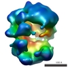 EMDB-1294: |
| Source |
|
 Homo sapiens (human)