 Movie
Movie Controller
 Structure viewers
 About Yorodumi Papers


+Search query
-Structure paper
| Title | A 3-D reconstruction of smooth muscle alpha-actinin by CryoEm reveals two different conformations at the actin-binding region. |
|---|---|
| Journal, issue, pages | J Mol Biol, Vol. 338, Issue 1, Page 115-125, Year 2004 |
| Publish date | Apr 16, 2004 |
 Authors Authors | Jun Liu / Dianne W Taylor / Kenneth A Taylor /  |
| PubMed Abstract | Cryoelectron microscopy was used to obtain a 3-D image at 2.0 nm resolution of 2-D arrays of smooth muscle alpha-actinin. The reconstruction reveals a well-resolved long central domain with 90 ...Cryoelectron microscopy was used to obtain a 3-D image at 2.0 nm resolution of 2-D arrays of smooth muscle alpha-actinin. The reconstruction reveals a well-resolved long central domain with 90 degrees of left-handed twist and near 2-fold symmetry. However, the molecular ends which contain the actin binding and calmodulin-like domains, have different structures oriented approximately 90 degrees to each other. Atomic structures for the alpha-actinin domains were built by homology modeling and assembled into an atomic model. Model building suggests that in the 2-D arrays, the two calponin homology domains that comprise the actin-binding domain have a closed conformation at one end and an open conformation at the other end due to domain swapping. The open and closed conformations of the actin-binding domain suggests flexibility that may underlie Ca2+ regulation. The approximately 90 degrees orientation difference at the molecular ends may underlie alpha-actinin's ability to crosslink actin filaments in nearly any orientation. |
 External links External links | J Mol Biol / PubMed:15050827 |
| Methods | EM (electron crystallography) |
| Resolution | 20 Å |
| Structure data | 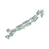 PDB-1sjj: |
| Source |
|
 Keywords Keywords | CONTRACTILE PROTEIN / 3-helix bundle / calponin homology domain / calmodulin-like domain / actin binding protein |

