 ムービー
ムービー コントローラー
 構造ビューア
 万見文献について


+検索条件
-Structure paper
| タイトル | Three-dimensional structure of a voltage-gated potassium channel at 2.5 nm resolution. |
|---|---|
| ジャーナル・号・ページ | Structure, Vol. 9, Issue 3, Page 215-220, Year 2001 |
| 掲載日 | 2001年3月7日 |
 著者 著者 | O Sokolova / L Kolmakova-Partensky / N Grigorieff /  |
| PubMed 要旨 | BACKGROUND: The voltage-gated potassium channel Shaker from Drosophila consists of a tetramer of identical subunits, each containing six transmembrane segments. The atomic structure of a bacterial ...BACKGROUND: The voltage-gated potassium channel Shaker from Drosophila consists of a tetramer of identical subunits, each containing six transmembrane segments. The atomic structure of a bacterial homolog, the potassium channel KcsA, is much smaller than Shaker. It does not have a voltage sensor and other important domains like the N-terminal tetramerization (T1) domain. The structure of these additional elements has to be studied in the more complex voltage-gated channels. RESULTS: We determined the three-dimensional structure of the entire Shaker channel at 2.5 nm resolution using electron microscopy. The four-fold symmetric structure shows a large and a small domain linked by thin 2 nm long connectors. To interpret the structure, we used the crystal structures of the isolated T1 domain and the KcsA channel. A unique density assignment was made based on the symmetry and dimensions of the crystal structures and domains, identifying the smaller domain as the cytoplasmic mass of Shaker containing T1 and the larger domain as embedded in the membrane. CONCLUSIONS: The two-domain architecture of the Shaker channel is consistent with the recently proposed "hanging gondola" model for the T1 domain, putting the T1 domain at a distance from the membrane domain but attached to it by thin connectors. The space between the two domains is sufficient to permit cytoplasmic access of ions and the N-terminal inactivation domain to the pore region. A hanging gondola architecture has also been observed in the nicotinic acetylcholine receptor and the KcsA structure, suggesting that it is a common element of ion channels. |
 リンク リンク | Structure / PubMed:11286888 |
| 手法 | EM (単粒子) |
| 解像度 | 25.0 Å |
| 構造データ | 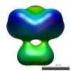 EMDB-1367: |
| 由来 |
|

