 ムービー
ムービー コントローラー
 構造ビューア
 EMN検索について


-検索条件
-検索結果
検索 (著者・登録者: brown & ii & ba)の結果全33件を表示しています

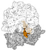
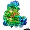
EMDB-19067: 
Cryo-EM structure of P. urativorans 70S ribosome in complex with hibernation factors Balon and RaiA (structure 1).

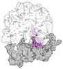
EMDB-19076:
Cryo-EM structure of P. urativorans 70S ribosome in complex with hibernation factor Balon, mRNA and P-site tRNA (structure 2).

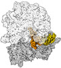
EMDB-19077:
Cryo-EM structure of P. urativorans 70S ribosome in complex with hibernation factor Balon and EF-Tu(GDP) (structure 3).

PDB-8rd8:
Cryo-EM structure of P. urativorans 70S ribosome in complex with hibernation factors Balon and RaiA (structure 1).

PDB-8rdv:
Cryo-EM structure of P. urativorans 70S ribosome in complex with hibernation factor Balon, mRNA and P-site tRNA (structure 2).

PDB-8rdw:
Cryo-EM structure of P. urativorans 70S ribosome in complex with hibernation factor Balon and EF-Tu(GDP) (structure 3).
EMDB-43074:
Cryo-EM structure of the Mycobacterium smegmatis 70S ribosome in complex with hibernation factor Msmeg1130 (Balon) (Structure 4)
EMDB-43075:
Cryo-EM structure of the Mycobacterium smegmatis 70S ribosome in complex with hibernation factor Rv2629 (Balon) (Structure 5)
EMDB-43076:
Cryo-EM structure of the Mycobacterium smegmatis 70S ribosome in complex with hibernation factor Msmeg1130 (Balon) and MsmegEF-Tu(GDP) (Composite structure 6)

EMDB-43077:
Cryo-EM structure of the Mycobacterium smegmatis 70S ribosome in complex with hibernation factor Msmeg1130 (Balon) and MsmegEF-Tu(GDP) (Structure 6)

EMDB-43078:
Hibernation factor Msmeg1130 (Balon) and MsmegEF-Tu(GDP) bound to Mycobacterium smegmatis 70S ribosome, from focused 3D classification and refinement (Structure 6)

PDB-8v9j:
Cryo-EM structure of the Mycobacterium smegmatis 70S ribosome in complex with hibernation factor Msmeg1130 (Balon) (Structure 4)

PDB-8v9k:
Cryo-EM structure of the Mycobacterium smegmatis 70S ribosome in complex with hibernation factor Rv2629 (Balon) (Structure 5)

PDB-8v9l:
Cryo-EM structure of the Mycobacterium smegmatis 70S ribosome in complex with hibernation factor Msmeg1130 (Balon) and MsmegEF-Tu(GDP) (Composite structure 6)
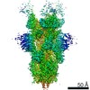
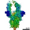
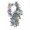
EMDB-23693:
Structure of the SARS-CoV-2 S 6P trimer in complex with the human neutralizing antibody Fab fragment, BG10-19
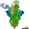
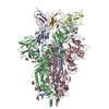
EMDB-23694:
Structure of the SARS-CoV-2 S 6P trimer in complex with the human neutralizing antibody Fab fragment, BG1-22
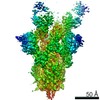
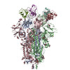
EMDB-23695:
Structure of the SARS-CoV-2 S 6P trimer in complex with the human neutralizing antibody Fab fragment, BG7-15

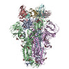
EMDB-23696:
Structure of the SARS-CoV-2 S 2P trimer in complex with the human neutralizing antibody Fab fragment, BG7-20
EMDB-23697:
Structure of the SARS-CoV-2 S 2P trimer in complex with the human neutralizing antibody Fab fragment, BG1-24
PDB-7m6e:
Structure of the SARS-CoV-2 S 6P trimer in complex with the human neutralizing antibody Fab fragment, BG10-19
PDB-7m6f:
Structure of the SARS-CoV-2 S 6P trimer in complex with the human neutralizing antibody Fab fragment, BG1-22

PDB-7m6g:
Structure of the SARS-CoV-2 S 6P trimer in complex with the human neutralizing antibody Fab fragment, BG7-15
PDB-7m6h:
Structure of the SARS-CoV-2 S 2P trimer in complex with the human neutralizing antibody Fab fragment, BG7-20
PDB-7m6i:
Structure of the SARS-CoV-2 S 2P trimer in complex with the human neutralizing antibody Fab fragment, BG1-24
EMDB-2929:
Mechanism of Ubiquitin Ligation to a Substrate by human Anaphase-Promoting Complex

PDB-4d3e:
Tetramer of IpaD, modified from 2J0O, fitted into negative stain electron microscopy reconstruction of the wild type tip complex from the type III secretion system of Shigella flexneri
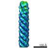
EMDB-2801:
Negative stain electron microscopy reconstruction of the wild type tip complex from the type III secretion system of Shigella flexneri
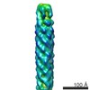
EMDB-2802:
Negative stain electron microscopy reconstruction of the tip complex from the type III secretion system of Shigella flexneri lacking IpaB
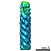
EMDB-2803:
Negative stain electron microscopy reconstruction of the tip complex and needle from the type III secretion system of Shigella flexneri with MxiH mutation P44A
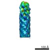
EMDB-2805:
Refined negative stain electron microscopy reconstruction of the tip complex from the type III secretion system of Shigella flexneri with MxiH mutation Q51A

EMDB-2806:
Negative stain electron microscopy reconstruction of the tip complex from the type III secretion system of Shigella flexneri with MxiH mutations P44A+Q51A

EMDB-2804:
Negative stain electron microscopy reconstruction of the tip complex from the type III secretion system of Shigella flexneri with MxiH mutation Q51A
EMDB-1636:
A dimeric structure for archaeal methylation-guide small ribonucleoproteins
 wwPDBはEMDBデータモデルのバージョン3へ移行します
