Filamentous archaeal viruses coat protein, C-terminal / helical viral capsid / DNA binding / DNA / DNA (> 10) / DNA (> 100) / Major capsid protein 1 / Major capsid protein 2
Function and homology information
Biological species
Acidianus filamentous virus 1
Method
ELECTRON MICROSCOPY / helical reconstruction / cryo EM / Resolution: 4.5 Å
National Institutes of Health/National Institute of General Medical Sciences (NIH/NIGMS)
GM035269
United States
National Institutes of Health/National Institute of General Medical Sciences (NIH/NIGMS)
GM098304
United States
French National Research Agency
ANR-13-BSV3-0017-01
France
Citation
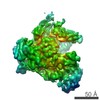
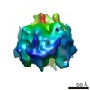
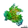
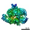
Journal: Elife / Year: 2017 Title: Model for a novel membrane envelope in a filamentous hyperthermophilic virus. Authors: Peter Kasson / Frank DiMaio / Xiong Yu / Soizick Lucas-Staat / Mart Krupovic / Stefan Schouten / David Prangishvili / Edward H Egelman / Abstract: Biological membranes create compartments, and are usually formed by lipid bilayers. However, in hyperthermophilic archaea that live optimally at temperatures above 80°C the membranes are monolayers ...Biological membranes create compartments, and are usually formed by lipid bilayers. However, in hyperthermophilic archaea that live optimally at temperatures above 80°C the membranes are monolayers which resemble fused bilayers. Many double-stranded DNA viruses which parasitize such hosts, including the filamentous virus AFV1 of , are enveloped with a lipid-containing membrane. Using cryo-EM, we show that the membrane in AFV1 is a ~2 nm-thick monolayer, approximately half the expected membrane thickness, formed by host membrane-derived lipids which adopt a U-shaped 'horseshoe' conformation. We hypothesize that this unusual viral envelope structure results from the extreme curvature of the viral capsid, as 'horseshoe' lipid conformations favor such curvature and host membrane lipids that permit horseshoe conformations are selectively recruited into the viral envelope. The unusual envelope found in AFV1 also has many implications for biotechnology, since this membrane can survive the most aggressive conditions involving extremes of temperature and pH.
Helical symmetry: (Circular symmetry: 1 / Dyad axis: no / N subunits divisor: 1 / Num. of operations: 30 / Rise per n subunits: 4.6 Å / Rotation per n subunits: 38.7 °)
-
Components
#1: Protein
... ORF140
Mass: 15832.856 Da / Num. of mol.: 21 / Source method: isolated from a natural source / Source: (natural) Acidianus filamentous virus 1 / References: UniProt: Q70LC6
#2: Protein
... ORF132
Mass: 15017.159 Da / Num. of mol.: 21 / Source method: isolated from a natural source / Source: (natural) Acidianus filamentous virus 1 / References: UniProt: Q70LC7
#3: DNA chain
DNA (253-MER)
Mass: 77747.367 Da / Num. of mol.: 2 / Source method: isolated from a natural source / Source: (natural) Acidianus filamentous virus 1
-
Experimental details
-
Experiment
Experiment
Method: ELECTRON MICROSCOPY
EM experiment
Aggregation state: FILAMENT / 3D reconstruction method: helical reconstruction
In the structure databanks used in Yorodumi, some data are registered as the other names, "COVID-19 virus" and "2019-nCoV". Here are the details of the virus and the list of structure data.
Jan 31, 2019. EMDB accession codes are about to change! (news from PDBe EMDB page)
EMDB accession codes are about to change! (news from PDBe EMDB page)
The allocation of 4 digits for EMDB accession codes will soon come to an end. Whilst these codes will remain in use, new EMDB accession codes will include an additional digit and will expand incrementally as the available range of codes is exhausted. The current 4-digit format prefixed with “EMD-” (i.e. EMD-XXXX) will advance to a 5-digit format (i.e. EMD-XXXXX), and so on. It is currently estimated that the 4-digit codes will be depleted around Spring 2019, at which point the 5-digit format will come into force.
The EM Navigator/Yorodumi systems omit the EMD- prefix.
Related info.:Q: What is EMD? / ID/Accession-code notation in Yorodumi/EM Navigator
Yorodumi is a browser for structure data from EMDB, PDB, SASBDB, etc.
This page is also the successor to EM Navigator detail page, and also detail information page/front-end page for Omokage search.
The word "yorodu" (or yorozu) is an old Japanese word meaning "ten thousand". "mi" (miru) is to see.
Related info.:EMDB / PDB / SASBDB / Comparison of 3 databanks / Yorodumi Search / Aug 31, 2016. New EM Navigator & Yorodumi / Yorodumi Papers / Jmol/JSmol / Function and homology information / Changes in new EM Navigator and Yorodumi
 Movie
Movie Controller

 Yorodumi
 Open data
 Basic information
 Components
 Keywords
 VIRUS / AFV1 /
  Function and homology information
 Authors
 United States,
  France, 3items
  Citation
 Structure visualization
 Downloads & links
 Other downloads


 PDBj
PDBj


 Assembly
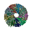
 Sample preparation
 Electron microscopy imaging
 Processing