 Movie
Movie Controller

[English] 日本語
 Yorodumi
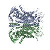
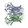
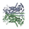
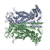
- PDB-5oyg: Structure of calcium-free mTMEM16A chloride channel at 4.06 A res... -
+ Open data

- Basic information
| Entry | Database: PDB / ID: 5oyg | |||||||||
|---|---|---|---|---|---|---|---|---|---|---|
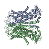
| Title | Structure of calcium-free mTMEM16A chloride channel at 4.06 A resolution | |||||||||
 Components Components | Anoctamin-1 Calcium-dependent chloride channel Calcium-dependent chloride channel | |||||||||
 Keywords Keywords | MEMBRANE PROTEIN / TMEM16 family / ion channel / cryo-EM | |||||||||
| Function / homology |  Function and homology information Function and homology informationglial cell projection elongation / trachea development / iodide transmembrane transporter activity / iodide transport / intracellularly calcium-gated chloride channel activity / mucus secretion / cellular response to peptide / Stimuli-sensing channels / voltage-gated chloride channel activity / calcium-activated cation channel activity ...glial cell projection elongation / trachea development / iodide transmembrane transporter activity / iodide transport / intracellularly calcium-gated chloride channel activity / mucus secretion / cellular response to peptide / Stimuli-sensing channels / voltage-gated chloride channel activity / calcium-activated cation channel activity / protein localization to membrane / chloride transport / chloride channel activity / positive regulation of insulin secretion involved in cellular response to glucose stimulus / detection of temperature stimulus involved in sensory perception of pain / chloride channel complex / monoatomic cation transport / chloride transmembrane transport / regulation of membrane potential / cell projection / establishment of localization in cell / presynaptic membrane / cellular response to heat / phospholipase C-activating G protein-coupled receptor signaling pathway / apical plasma membrane / external side of plasma membrane / signaling receptor binding / glutamatergic synapse / protein homodimerization activity / nucleoplasm / identical protein binding / metal ion binding / plasma membraneSimilarity search - Function | |||||||||
| Biological species |  Mus musculus (house mouse) Mus musculus (house mouse) | |||||||||
| Method | ELECTRON MICROSCOPY / single particle reconstruction / cryo EM / Resolution: 4.06 Å | |||||||||
 Authors Authors | Paulino, C. / Kalienkova, V. / Lam, K.M. / Neldner, Y. / Dutzler, R. | |||||||||
| Funding support |  Switzerland, 2items Switzerland, 2items
| |||||||||
 Citation Citation | Journal: Nature / Year: 2017 Title: Activation mechanism of the calcium-activated chloride channel TMEM16A revealed by cryo-EM. Authors: Cristina Paulino / Valeria Kalienkova / Andy K M Lam / Yvonne Neldner / Raimund Dutzler / Abstract: The calcium-activated chloride channel TMEM16A is a ligand-gated anion channel that opens in response to an increase in intracellular Ca concentration. The protein is broadly expressed and ...The calcium-activated chloride channel TMEM16A is a ligand-gated anion channel that opens in response to an increase in intracellular Ca concentration. The protein is broadly expressed and contributes to diverse physiological processes, including transepithelial chloride transport and the control of electrical signalling in smooth muscles and certain neurons. As a member of the TMEM16 (or anoctamin) family of membrane proteins, TMEM16A is closely related to paralogues that function as scramblases, which facilitate the bidirectional movement of lipids across membranes. The unusual functional diversity of the TMEM16 family and the relationship between two seemingly incompatible transport mechanisms has been the focus of recent investigations. Previous breakthroughs were obtained from the X-ray structure of the lipid scramblase of the fungus Nectria haematococca (nhTMEM16), and from the cryo-electron microscopy structure of mouse TMEM16A at 6.6 Å (ref. 14). Although the latter structure disclosed the architectural differences that distinguish ion channels from lipid scramblases, its low resolution did not permit a detailed molecular description of the protein or provide any insight into its activation by Ca. Here we describe the structures of mouse TMEM16A at high resolution in the presence and absence of Ca. These structures reveal the differences between ligand-bound and ligand-free states of a calcium-activated chloride channel, and when combined with functional experiments suggest a mechanism for gating. During activation, the binding of Ca to a site located within the transmembrane domain, in the vicinity of the pore, alters the electrostatic properties of the ion conduction path and triggers a conformational rearrangement of an α-helix that comes into physical contact with the bound ligand, and thereby directly couples ligand binding and pore opening. Our study describes a process that is unique among channel proteins, but one that is presumably general for both functional branches of the TMEM16 family. | |||||||||
| History |
|
- Structure visualization
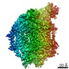
| Movie |
Movie viewer |
|---|---|
| Structure viewer | Molecule: MolmilJmol/JSmol |
- Downloads & links
-Download
| PDBx/mmCIF format | 5oyg.cif.gz | 297.1 KB | Display | PDBx/mmCIF format |
|---|---|---|---|---|
| PDB format | pdb5oyg.ent.gz | 243.2 KB | Display | PDB format |
| PDBx/mmJSON format | 5oyg.json.gz | Tree view | PDBx/mmJSON format | |
| Others |  Other downloads Other downloads |
-Validation report
| Arichive directory | https://data.pdbj.org/pub/pdb/validation_reports/oy/5oygftp://data.pdbj.org/pub/pdb/validation_reports/oy/5oyg | HTTPS FTP |
|---|
-Related structure data
| Related structure data |  3861MC  3860C  5oybC C: citing same article ( M: map data used to model this data |
|---|---|
| Similar structure data |


-Links
 PDBj
PDBj- Assembly
| Deposited unit | 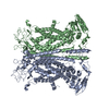
|
|---|---|
| 1 |
|
-Components
| #1: Protein | Calcium-dependent chloride channel / Transmembrane protein 16A Mass: 111058.992 Da / Num. of mol.: 2 Source method: isolated from a genetically manipulated source Source: (gene. exp.) Mus musculus (house mouse) / Gene: Ano1, Tmem16a / Cell line (production host): HEK293 / Production host:  Homo sapiens (human) / References: UniProt: Q8BHY3 Homo sapiens (human) / References: UniProt: Q8BHY3 |
|---|
-Experimental details
-Experiment
| Experiment | Method: ELECTRON MICROSCOPY |
|---|---|
| EM experiment | Aggregation state: PARTICLE / 3D reconstruction method: single particle reconstruction |
- Sample preparation
| Component | Name: mTMEM16A in absence of calcium ions / Type: COMPLEX / Details: calcium-activated chloride channel / Entity ID: all / Source: RECOMBINANT |
|---|---|
| Molecular weight | Value: 0.110916 MDa / Experimental value: YES |
| Source (natural) | Organism: Mus musculus (house mouse) |
| Source (recombinant) | Organism: Homo sapiens (human) / Strain: HEK293 / Cell: stabel mTMEM16A cell line (Flp-In System) |
| Buffer solution | pH: 7.5 / Details: 20 mM Hepes 150 mM NaCl <0.12% digitonin |
| Buffer component | Conc.: 20 mM / Name: Hepes |
| Specimen | Conc.: 3.3 mg/ml / Embedding applied: NO / Shadowing applied: NO / Staining applied: NO / Vitrification applied: YES Details: full-length (wild-type isoform ac) deglycosylated mTMEM16A in absence of CaCl2 |
| Specimen support | Grid material: GOLD / Grid mesh size: 200 divisions/in. / Grid type: Quantifoil R1.2/1.3 |
| Vitrification | Instrument: FEI VITROBOT MARK IV / Cryogen name: ETHANE / Humidity: 100 % / Chamber temperature: 288 K / Details: 2 ul sample volume 2-4 sec blotting time |
- Electron microscopy imaging
| Experimental equipment |  Model: Titan Krios / Image courtesy: FEI Company |
|---|---|
| Microscopy | Model: FEI TITAN KRIOS |
| Electron gun | Electron source: FIELD EMISSION GUN / Accelerating voltage: 300 kV / Illumination mode: FLOOD BEAM |
| Electron lens | Mode: BRIGHT FIELDBright-field microscopy / Nominal magnification: 36630 X / Calibrated magnification: 36630 X / Nominal defocus max: 3800 nm / Nominal defocus min: 500 nm / Calibrated defocus min: 500 nm / Calibrated defocus max: 3800 nm / Cs: 2.7 mm / C2 aperture diameter: 100 µm / Alignment procedure: COMA FREE |
| Specimen holder | Cryogen: NITROGEN / Specimen holder model: FEI TITAN KRIOS AUTOGRID HOLDER / Temperature (max): 100 K / Temperature (min): 80 K |
| Image recording | Average exposure time: 15 sec. / Electron dose: 80 e/Å2 / Detector mode: SUPER-RESOLUTION / Film or detector model: GATAN K2 SUMMIT (4k x 4k) / Num. of grids imaged: 3 / Num. of real images: 5063 Details: Data were collected in an automated fashion using SerialEM47 on a K2 Summit detector (Gatan). The dataset in absence of calcium ions was obtained at a pixel size of 0.6825A in super- ...Details: Data were collected in an automated fashion using SerialEM47 on a K2 Summit detector (Gatan). The dataset in absence of calcium ions was obtained at a pixel size of 0.6825A in super-resolution mode, a defocus range of -0.5 to -3.9 um, an exposure time of 15 sec and a sub-frame exposure time of 150 ms (100 frames) with an electron dose at the specimen level of 0.75-0.8 e-/A2/frame. The total accumulated dose on the specimen level was approximately 80 e-/A2. |
| EM imaging optics | Energyfilter name: In-column Omega Filter / Energyfilter upper: 10 eV / Energyfilter lower: -10 eV / Energyfilter slit width: 20 eV |
| Image scans | Sampling size: 5 µm / Width: 7420 / Height: 7676 / Movie frames/image: 100 / Used frames/image: 1-100 |
- Processing
| Software | Name: REFMAC / Version: 5.8.0158 / Classification: refinement | ||||||||||||||||||||||||||||||||||||||||||||||||||||||||||||||||||||||||||||||||||||||||||||||||||||||||||
|---|---|---|---|---|---|---|---|---|---|---|---|---|---|---|---|---|---|---|---|---|---|---|---|---|---|---|---|---|---|---|---|---|---|---|---|---|---|---|---|---|---|---|---|---|---|---|---|---|---|---|---|---|---|---|---|---|---|---|---|---|---|---|---|---|---|---|---|---|---|---|---|---|---|---|---|---|---|---|---|---|---|---|---|---|---|---|---|---|---|---|---|---|---|---|---|---|---|---|---|---|---|---|---|---|---|---|---|
| EM software |
| ||||||||||||||||||||||||||||||||||||||||||||||||||||||||||||||||||||||||||||||||||||||||||||||||||||||||||
| Image processing | Details: Fourier cropping (final pixel size 1.365 A), motion correction and dose-weighting of frames were performed by MotionCor2 | ||||||||||||||||||||||||||||||||||||||||||||||||||||||||||||||||||||||||||||||||||||||||||||||||||||||||||
| CTF correction | Details: The contrast transfer function (CTF) parameters were estimated on the movie frames by ctffind4.1 Type: PHASE FLIPPING AND AMPLITUDE CORRECTION | ||||||||||||||||||||||||||||||||||||||||||||||||||||||||||||||||||||||||||||||||||||||||||||||||||||||||||
| Particle selection | Num. of particles selected: 1349821 Details: For the dataset collected in absence of calcium ions a total of 5063 dose-fractionated super-resolution images were recorded, 2 x 2 down-sampled by Fourier cropping (final pixel size 1.365A) ...Details: For the dataset collected in absence of calcium ions a total of 5063 dose-fractionated super-resolution images were recorded, 2 x 2 down-sampled by Fourier cropping (final pixel size 1.365A) and subjected to motion correction and dose-weighting of frames by MotionCor2. The contrast transfer function (CTF) parameters were estimated on the movie frames by ctffind4.1. Images showing a strong drift, higher defocus than -3.8 um or a bad CTF estimation were discarded, From a total of 4,738 images (final pixel size 1.365 A) 1,349,821 particles were extracted with a box size of 256 pixels after auto-picking and initial round of 2D classification. Initial rounds of 2D and 3D classification, resulted in a set of 467,286 particles which where subjected to particle polishing, as described above, as well as final rounds of 3D classification. The final polished and auto-refined map was calculated from 195,465 particles derived from 4726 images with a resolution of 4.86 A before masking and 4.06 A after masking and was sharpened using an isotropic b-factor of -116 A2 | ||||||||||||||||||||||||||||||||||||||||||||||||||||||||||||||||||||||||||||||||||||||||||||||||||||||||||
| 3D reconstruction | Resolution: 4.06 Å / Resolution method: FSC 0.143 CUT-OFF / Num. of particles: 195465 / Symmetry type: POINT | ||||||||||||||||||||||||||||||||||||||||||||||||||||||||||||||||||||||||||||||||||||||||||||||||||||||||||
| Atomic model building | Protocol: RIGID BODY FIT | ||||||||||||||||||||||||||||||||||||||||||||||||||||||||||||||||||||||||||||||||||||||||||||||||||||||||||
| Refinement | Resolution: 4.06→120.12 Å / Cor.coef. Fo:Fc: 0.519 Stereochemistry target values: MAXIMUM LIKELIHOOD WITH PHASES Details: HYDROGENS HAVE BEEN ADDED IN THE RIDING POSITIONS
| ||||||||||||||||||||||||||||||||||||||||||||||||||||||||||||||||||||||||||||||||||||||||||||||||||||||||||
| Solvent computation | Ion probe radii: 0.8 Å / Shrinkage radii: 0.8 Å / VDW probe radii: 1.2 Å / Solvent model: MASK | ||||||||||||||||||||||||||||||||||||||||||||||||||||||||||||||||||||||||||||||||||||||||||||||||||||||||||
| Displacement parameters | Biso mean: 161.117 Å2
| ||||||||||||||||||||||||||||||||||||||||||||||||||||||||||||||||||||||||||||||||||||||||||||||||||||||||||
| Refinement step | Cycle: 1 / Total: 11596 | ||||||||||||||||||||||||||||||||||||||||||||||||||||||||||||||||||||||||||||||||||||||||||||||||||||||||||
| Refine LS restraints |
|