 Movie
Movie Controller

[English] 日本語
 Yorodumi
- EMDB-3403: Asymmetric cryo-EM reconstruction of phage MS2 reveals genome str... -
+ Open data

- Basic information
| Entry | Database: EMDB / ID: EMD-3403 | |||||||||
|---|---|---|---|---|---|---|---|---|---|---|
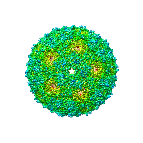
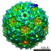
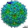
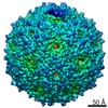
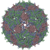
| Title | Asymmetric cryo-EM reconstruction of phage MS2 reveals genome structure in situ | |||||||||
 Map data Map data | Asymmetric reconstruction of MS2 virion | |||||||||
 Sample Sample |
| |||||||||
 Keywords Keywords | MS2 / RNA genome / structure / cryo-EM | |||||||||
| Biological species |  Enterobacterio phage MS2 (virus) Enterobacterio phage MS2 (virus) | |||||||||
| Method | single particle reconstruction / cryo EM / negative staining / Resolution: 10.5 Å | |||||||||
 Authors Authors | Koning RI / Gomez-Blanco J / Akopjana I / Vargas J / Kazaks A / Tars K / Carazo JM / Koster AJ | |||||||||
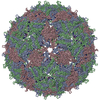
 Citation Citation | Journal: Nat Commun / Year: 2016 Title: Asymmetric cryo-EM reconstruction of phage MS2 reveals genome structure in situ. Authors: Roman I Koning / Josue Gomez-Blanco / Inara Akopjana / Javier Vargas / Andris Kazaks / Kaspars Tars / José María Carazo / Abraham J Koster /    Abstract: In single-stranded ribonucleic acid (RNA) viruses, virus capsid assembly and genome packaging are intertwined processes. Using cryo-electron microscopy and single particle analysis we determined the ...In single-stranded ribonucleic acid (RNA) viruses, virus capsid assembly and genome packaging are intertwined processes. Using cryo-electron microscopy and single particle analysis we determined the asymmetric virion structure of bacteriophage MS2, which includes 178 copies of the coat protein, a single copy of the A-protein and the RNA genome. This reveals that in situ, the viral RNA genome can adopt a defined conformation. The RNA forms a branched network of stem-loops that almost all allocate near the capsid inner surface, while predominantly binding to coat protein dimers that are located in one-half of the capsid. This suggests that genomic RNA is highly involved in genome packaging and virion assembly. | |||||||||
| History |
|
- Structure visualization
| Movie |
 Movie viewer Movie viewer |
|---|---|
| Structure viewer | EM map: SurfViewMolmilJmol/JSmol |
| Supplemental images |
- Downloads & links
-EMDB archive
| Map data | emd_3403.map.gz | 105.6 MB | EMDB map data format | |
|---|---|---|---|---|
| Header (meta data) | emd-3403-v30.xmlemd-3403.xml | 10.6 KB 10.6 KB | Display Display | EMDB header |
| Images |  EMD-3403.png EMD-3403.png | 198.7 KB | ||
| Archive directory |  http://ftp.pdbj.org/pub/emdb/structures/EMD-3403ftp://ftp.pdbj.org/pub/emdb/structures/EMD-3403 http://ftp.pdbj.org/pub/emdb/structures/EMD-3403ftp://ftp.pdbj.org/pub/emdb/structures/EMD-3403 | HTTPS FTP |
-Validation report
| Summary document | emd_3403_validation.pdf.gz | 304.9 KB | Display | EMDB validaton report |
|---|---|---|---|---|
| Full document | emd_3403_full_validation.pdf.gz | 304 KB | Display | |
| Data in XML | emd_3403_validation.xml.gz | 6.6 KB | Display | |
| Arichive directory | https://ftp.pdbj.org/pub/emdb/validation_reports/EMD-3403ftp://ftp.pdbj.org/pub/emdb/validation_reports/EMD-3403 | HTTPS FTP |
-Related structure data
| Related structure data |  3402C  3404C  3540C C: citing same article ( |
|---|---|
| Similar structure data | |
| EM raw data | EMPIAR-10075 (Title: Asymmetric cryo-EM reconstruction of phage MS2 reveals genome structure in situ Data size: 46.9 Data #1: Aligned 7-frame micrographs of bacteriophage MS2. [micrographs - single frame]) |


-Links
| EMDB pages | EMDB (EBI/PDBe) / EMDataResource |
|---|
-Map
| File | Download / File: emd_3403.map.gz / Format: CCP4 / Size: 111 MB / Type: IMAGE STORED AS FLOATING POINT NUMBER (4 BYTES) | ||||||||||||||||||||||||||||||||||||||||||||||||||||||||||||||||||||
|---|---|---|---|---|---|---|---|---|---|---|---|---|---|---|---|---|---|---|---|---|---|---|---|---|---|---|---|---|---|---|---|---|---|---|---|---|---|---|---|---|---|---|---|---|---|---|---|---|---|---|---|---|---|---|---|---|---|---|---|---|---|---|---|---|---|---|---|---|---|
| Annotation | Asymmetric reconstruction of MS2 virion | ||||||||||||||||||||||||||||||||||||||||||||||||||||||||||||||||||||
| Voxel size | X=Y=Z: 1.14 Å | ||||||||||||||||||||||||||||||||||||||||||||||||||||||||||||||||||||
| Density |
| ||||||||||||||||||||||||||||||||||||||||||||||||||||||||||||||||||||
| Symmetry | Space group: 1 | ||||||||||||||||||||||||||||||||||||||||||||||||||||||||||||||||||||
| Details | EMDB XML:
CCP4 map header:
| ||||||||||||||||||||||||||||||||||||||||||||||||||||||||||||||||||||
-Supplemental data
- Sample components
-Entire : Bacteriophage MS2
| Entire | Name: Bacteriophage MS2 (virus) |
|---|---|
| Components |
|
-Supramolecule #1000: Bacteriophage MS2
| Supramolecule | Name: Bacteriophage MS2 / type: sample / ID: 1000 / Details: Purified by gel filtration Oligomeric state: One copy of genomic RNA inside a shell of 178 capsid proteins and one A-protein Number unique components: 1 |
|---|---|
| Molecular weight | Theoretical: 3.6 MDa |
-Supramolecule #1: Enterobacterio phage MS2
| Supramolecule | Name: Enterobacterio phage MS2 / type: virus / ID: 1 / Name.synonym: bacteriophage MS2 / NCBI-ID: 12022 / Sci species name: Enterobacterio phage MS2 / Virus type: VIRION / Virus isolate: SUBSPECIES / Virus enveloped: No / Virus empty: No / Syn species name: bacteriophage MS2 / Sci species subspecies: Enterobacterio phage MS2 |
|---|---|
| Host (natural) | Organism:  |
| Molecular weight | Theoretical: 3.6 MDa |
| Virus shell | Shell ID: 1 / Name: MS2 / Diameter: 275 Å / T number (triangulation number): 3 |
-Experimental details
-Structure determination
| Method | negative staining, cryo EM |
|---|---|
 Processing Processing | single particle reconstruction |
| Aggregation state | particle |
-Sample preparation
| Concentration | 1 mg/mL |
|---|---|
| Buffer | pH: 7.4 / Details: PBS |
| Staining | Type: NEGATIVE / Details: Vitrification |
| Grid | Details: Cu 300 Mesh with quantifoil R2/2 support film, glow discharged in air at 0.2 mbar for 1 minute at 30 mA. |
| Vitrification | Cryogen name: ETHANE-PROPANE MIXTURE / Chamber humidity: 70 % / Chamber temperature: 100 K / Instrument: LEICA EM GP Method: Blotted using filter paper for 1 to 2 seconds before blotting |
- Electron microscopy
| Microscope | FEI TITAN KRIOS |
|---|---|
| Temperature | Min: 77 K / Max: 97 K / Average: 87 K |
| Alignment procedure | Legacy - Astigmatism: Cs corrector / Legacy - Electron beam tilt params: 15 |
| Date | May 19, 2014 |
| Image recording | Category: CCD / Film or detector model: FEI FALCON II (4k x 4k) / Digitization - Sampling interval: 14 µm / Number real images: 751 / Average electron dose: 35 e/Å2 Details: Images are average of 7 frames recorded in movie mode on direct electron detector Bits/pixel: 16 |
| Electron beam | Acceleration voltage: 300 kV / Electron source:  FIELD EMISSION GUN FIELD EMISSION GUN |
| Electron optics | Calibrated magnification: 61403 / Illumination mode: FLOOD BEAM / Imaging mode: BRIGHT FIELD / Cs: 0.02 mm / Nominal defocus max: 2.5 µm / Nominal defocus min: 0.5 µm / Nominal magnification: 59000 |
| Sample stage | Specimen holder: Nitrogen cooled / Specimen holder model: FEI TITAN KRIOS AUTOGRID HOLDER |
| Experimental equipment |  Model: Titan Krios / Image courtesy: FEI Company |
-Image processing
| Details | Movies were aligned using Optical Flow, CTFs were estimated using CTFFIND3, and were used to select the best quality micrographs. A total of 22,441 particles were picked automatically using Xmipp. Particles were classified using 2D reference-free Relion approach. Relion was used for 3D refinement using icosahedral symmetry and gold-standard approach. |
|---|---|
| CTF correction | Details: Per image |
| Final reconstruction | Applied symmetry - Point group: C1 (asymmetric) / Algorithm: OTHER / Resolution.type: BY AUTHOR / Resolution: 10.5 Å / Resolution method: OTHER / Software - Name: Scipion, Xmipp, CTFFIND3, Relion / Number images used: 18977 |
| Final two d classification | Number classes: 5201 |
