 Movie
Movie Controller


+ Open data

- Basic information
| Entry | Database: EMDB / ID: EMD-1530 | |||||||||
|---|---|---|---|---|---|---|---|---|---|---|
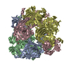
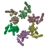
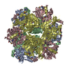
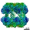
| Title | Structure of hexameric DyP from Brevibacterium linens | |||||||||
 Map data Map data | Structure of hexameric DyP from Brevibacterium linens | |||||||||
 Sample Sample |
| |||||||||
 Keywords Keywords | protein complex / hexamer / peroxidase / dye-decolorizing peroxidase / heme containing peroxidase | |||||||||
| Function / homology | Dyp-type peroxidase Function and homology information Function and homology information | |||||||||
| Biological species |  Brevibacterium linens (bacteria) Brevibacterium linens (bacteria) | |||||||||
| Method | single particle reconstruction / negative staining / Resolution: 16.0 Å | |||||||||
 Authors Authors | Sutter M / Boehringer D / Gutmann S / Gunther S / Prangishvili D / Loessner MJ / Stetter KO / Weber-Ban E / Ban N | |||||||||
 Citation Citation | Journal: Nat Struct Mol Biol / Year: 2008 Title: Structural basis of enzyme encapsulation into a bacterial nanocompartment. Authors: Markus Sutter / Daniel Boehringer / Sascha Gutmann / Susanne Günther / David Prangishvili / Martin J Loessner / Karl O Stetter / Eilika Weber-Ban / Nenad Ban /  Abstract: Compartmentalization is an important organizational feature of life. It occurs at varying levels of complexity ranging from eukaryotic organelles and the bacterial microcompartments, to the molecular ...Compartmentalization is an important organizational feature of life. It occurs at varying levels of complexity ranging from eukaryotic organelles and the bacterial microcompartments, to the molecular reaction chambers formed by enzyme assemblies. The structural basis of enzyme encapsulation in molecular compartments is poorly understood. Here we show, using X-ray crystallographic, biochemical and EM experiments, that a widespread family of conserved bacterial proteins, the linocin-like proteins, form large assemblies that function as a minimal compartment to package enzymes. We refer to this shell-forming protein as 'encapsulin'. The crystal structure of such a particle from Thermotoga maritima determined at 3.1-angstroms resolution reveals that 60 copies of the monomer assemble into a thin, icosahedral shell with a diameter of 240 angstroms. The interior of this nanocompartment is lined with conserved binding sites for short polypeptide tags present as C-terminal extensions of enzymes involved in oxidative-stress response. | |||||||||
| History |
|
- Structure visualization
| Movie |
Movie viewer |
|---|---|
| Structure viewer | EM map: SurfViewMolmilJmol/JSmol |
| Supplemental images |

- Downloads & links
-EMDB archive
| Map data | emd_1530.map.gz | 623.7 KB | EMDB map data format | |
|---|---|---|---|---|
| Header (meta data) | emd-1530-v30.xmlemd-1530.xml | 8.9 KB 8.9 KB | Display Display | EMDB header |
| Images |  1530.gif 1530.gif | 68.1 KB | ||
| Archive directory |  http://ftp.pdbj.org/pub/emdb/structures/EMD-1530ftp://ftp.pdbj.org/pub/emdb/structures/EMD-1530 http://ftp.pdbj.org/pub/emdb/structures/EMD-1530ftp://ftp.pdbj.org/pub/emdb/structures/EMD-1530 | HTTPS FTP |
-Validation report
| Summary document | emd_1530_validation.pdf.gz | 205.8 KB | Display | EMDB validaton report |
|---|---|---|---|---|
| Full document | emd_1530_full_validation.pdf.gz | 204.9 KB | Display | |
| Data in XML | emd_1530_validation.xml.gz | 4.8 KB | Display | |
| Arichive directory | https://ftp.pdbj.org/pub/emdb/validation_reports/EMD-1530ftp://ftp.pdbj.org/pub/emdb/validation_reports/EMD-1530 | HTTPS FTP |
-Related structure data


-Links
| EMDB pages | EMDB (EBI/PDBe) / EMDataResource |
|---|
-Map
| File | Download / File: emd_1530.map.gz / Format: CCP4 / Size: 1001 KB / Type: IMAGE STORED AS FLOATING POINT NUMBER (4 BYTES) | ||||||||||||||||||||||||||||||||||||||||||||||||||||||||||||||||||||
|---|---|---|---|---|---|---|---|---|---|---|---|---|---|---|---|---|---|---|---|---|---|---|---|---|---|---|---|---|---|---|---|---|---|---|---|---|---|---|---|---|---|---|---|---|---|---|---|---|---|---|---|---|---|---|---|---|---|---|---|---|---|---|---|---|---|---|---|---|---|
| Annotation | Structure of hexameric DyP from Brevibacterium linens | ||||||||||||||||||||||||||||||||||||||||||||||||||||||||||||||||||||
| Voxel size | X=Y=Z: 2.8 Å | ||||||||||||||||||||||||||||||||||||||||||||||||||||||||||||||||||||
| Density |
| ||||||||||||||||||||||||||||||||||||||||||||||||||||||||||||||||||||
| Symmetry | Space group: 1 | ||||||||||||||||||||||||||||||||||||||||||||||||||||||||||||||||||||
| Details | EMDB XML:
CCP4 map header:
| ||||||||||||||||||||||||||||||||||||||||||||||||||||||||||||||||||||
-Supplemental data
- Sample components
-Entire : Structure of hexameric DyP from Brevibacterium linens
| Entire | Name: Structure of hexameric DyP from Brevibacterium linens |
|---|---|
| Components |
|
-Supramolecule #1000: Structure of hexameric DyP from Brevibacterium linens
| Supramolecule | Name: Structure of hexameric DyP from Brevibacterium linens / type: sample / ID: 1000 / Details: monodisperse / Oligomeric state: hexamer / Number unique components: 1 |
|---|---|
| Molecular weight | Theoretical: 240 KDa |
-Macromolecule #1: dye-decolorizing peroxidase
| Macromolecule | Name: dye-decolorizing peroxidase / type: protein_or_peptide / ID: 1 / Name.synonym: Dyp / Number of copies: 6 / Oligomeric state: hexamer / Recombinant expression: Yes |
|---|---|
| Source (natural) | Organism: Brevibacterium linens (bacteria) / Strain: M18 |
| Molecular weight | Theoretical: 40 KDa |
| Recombinant expression | Organism: Escherichia coli Rosetta / Recombinant plasmid: pET21a |
| Sequence | InterPro: Dyp-type peroxidase |
-Experimental details
-Structure determination
| Method | negative staining |
|---|---|
 Processing Processing | single particle reconstruction |
| Aggregation state | particle |
-Sample preparation
| Staining | Type: NEGATIVE / Details: 2% w/v uranyl acetate |
|---|---|
| Grid | Details: carbon coated 200 mesh copper grid |
| Vitrification | Cryogen name: NONE / Instrument: OTHER |
- Electron microscopy
| Microscope | FEI TECNAI 20 |
|---|---|
| Temperature | Average: 295 K |
| Specialist optics | Energy filter - Name: Tridiem Gatan |
| Image recording | Category: CCD / Film or detector model: GATAN ULTRASCAN 1000 (2k x 2k) / Digitization - Sampling interval: 1.4 µm / Average electron dose: 15 e/Å2 |
| Electron beam | Acceleration voltage: 200 kV / Electron source:  FIELD EMISSION GUN FIELD EMISSION GUN |
| Electron optics | Calibrated magnification: 100000 / Illumination mode: FLOOD BEAM / Imaging mode: BRIGHT FIELD / Nominal defocus max: 1.5 µm / Nominal defocus min: 0.7 µm / Nominal magnification: 100000 |
| Sample stage | Specimen holder: Eucentric / Specimen holder model: OTHER |
-Image processing
| CTF correction | Details: Each image |
|---|---|
| Final reconstruction | Applied symmetry - Point group: D3 (2x3 fold dihedral) / Algorithm: OTHER / Resolution.type: BY AUTHOR / Resolution: 16.0 Å / Resolution method: OTHER / Software - Name: IMAGIC-5 / Number images used: 2878 |
