 Movie
Movie Controller

[English] 日本語
 Yorodumi
- EMDB-1252: Nucleotide-dependent conformational changes in the DnaA-like core... -
+ Open data

- Basic information
| Entry | Database: EMDB / ID: EMD-1252 | |||||||||
|---|---|---|---|---|---|---|---|---|---|---|
| Title | Nucleotide-dependent conformational changes in the DnaA-like core of the origin recognition complex. | |||||||||
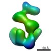
 Map data Map data | This is an envelope of the apo-form of the Drosophila melanogaster Origin Recognition Complex. | |||||||||
 Sample Sample |
| |||||||||
| Biological species |  | |||||||||
| Method | single particle reconstruction / cryo EM / Resolution: 34.0 Å | |||||||||
 Authors Authors | Clarey MG / Erzberger JP / Grob P / Leschziner AE / Berger JM / Nogales EN / Botchan MR | |||||||||
 Citation Citation | Journal: Nat Struct Mol Biol / Year: 2006 Title: Nucleotide-dependent conformational changes in the DnaA-like core of the origin recognition complex. Authors: Megan G Clarey / Jan P Erzberger / Patricia Grob / Andres E Leschziner / James M Berger / Eva Nogales / Michael Botchan /  Abstract: Structural details of initiator proteins for DNA replication have provided clues to the molecular events in this process. EM reconstructions of the Drosophila melanogaster origin recognition complex ...Structural details of initiator proteins for DNA replication have provided clues to the molecular events in this process. EM reconstructions of the Drosophila melanogaster origin recognition complex (ORC) reveal nucleotide-dependent conformational changes in the core of the complex. All five AAA+ domains in ORC contain a conserved structural element that, in DnaA, promotes formation of a right-handed helix, indicating that helical AAA+ substructures may be a feature of all initiators. A DnaA helical pentamer can be docked into ORC, and the location of Orc5 uniquely positions this core. The results suggest that ATP-dependent conformational changes observed in ORC derive from reorientation of the AAA+ domains. By analogy to the DNA-wrapping activity of DnaA, we posit that ORC together with Cdc6 prepares origin DNA for helicase loading through mechanisms related to the established pathway of prokaryotes. | |||||||||
| History |
|
- Structure visualization
| Movie |
 Movie viewer Movie viewer |
|---|---|
| Structure viewer | EM map: SurfViewMolmilJmol/JSmol |
| Supplemental images |
 UCSF Chimera
- Downloads & links
-EMDB archive
| Map data | emd_1252.map.gz | 1.2 MB | EMDB map data format | |
|---|---|---|---|---|
| Header (meta data) | emd-1252-v30.xmlemd-1252.xml | 7.1 KB 7.1 KB | Display Display | EMDB header |
| Images |  1252.gif 1252.gif | 20.8 KB | ||
| Archive directory |  http://ftp.pdbj.org/pub/emdb/structures/EMD-1252ftp://ftp.pdbj.org/pub/emdb/structures/EMD-1252 http://ftp.pdbj.org/pub/emdb/structures/EMD-1252ftp://ftp.pdbj.org/pub/emdb/structures/EMD-1252 | HTTPS FTP |
-Validation report
| Summary document | emd_1252_validation.pdf.gz | 224.3 KB | Display | EMDB validaton report |
|---|---|---|---|---|
| Full document | emd_1252_full_validation.pdf.gz | 223.4 KB | Display | |
| Data in XML | emd_1252_validation.xml.gz | 4.7 KB | Display | |
| Arichive directory | https://ftp.pdbj.org/pub/emdb/validation_reports/EMD-1252ftp://ftp.pdbj.org/pub/emdb/validation_reports/EMD-1252 | HTTPS FTP |
-Related structure data


-Links
| EMDB pages | EMDB (EBI/PDBe) / EMDataResource |
|---|
-Map
| File | Download / File: emd_1252.map.gz / Format: CCP4 / Size: 1.2 MB / Type: IMAGE STORED AS FLOATING POINT NUMBER (4 BYTES) | ||||||||||||||||||||||||||||||||||||||||||||||||||||||||||||||||||||
|---|---|---|---|---|---|---|---|---|---|---|---|---|---|---|---|---|---|---|---|---|---|---|---|---|---|---|---|---|---|---|---|---|---|---|---|---|---|---|---|---|---|---|---|---|---|---|---|---|---|---|---|---|---|---|---|---|---|---|---|---|---|---|---|---|---|---|---|---|---|
| Annotation | This is an envelope of the apo-form of the Drosophila melanogaster Origin Recognition Complex. | ||||||||||||||||||||||||||||||||||||||||||||||||||||||||||||||||||||
| Voxel size | X=Y=Z: 5.18 Å | ||||||||||||||||||||||||||||||||||||||||||||||||||||||||||||||||||||
| Density |
| ||||||||||||||||||||||||||||||||||||||||||||||||||||||||||||||||||||
| Symmetry | Space group: 1 | ||||||||||||||||||||||||||||||||||||||||||||||||||||||||||||||||||||
| Details | EMDB XML:
CCP4 map header:
| ||||||||||||||||||||||||||||||||||||||||||||||||||||||||||||||||||||
-Supplemental data
- Sample components
-Entire : Drosophila melanogaster Origin Recognition Complex
| Entire | Name: Drosophila melanogaster Origin Recognition Complex |
|---|---|
| Components |
|
-Supramolecule #1000: Drosophila melanogaster Origin Recognition Complex
| Supramolecule | Name: Drosophila melanogaster Origin Recognition Complex / type: sample / ID: 1000 / Number unique components: 1 |
|---|---|
| Molecular weight | Experimental: 420 KDa / Theoretical: 420 KDa |
-Macromolecule #1: Drosophila melanogaster Origin Recognition Complex
| Macromolecule | Name: Drosophila melanogaster Origin Recognition Complex / type: protein_or_peptide / ID: 1 / Name.synonym: ORC / Number of copies: 1 / Oligomeric state: heterohexamer / Recombinant expression: Yes |
|---|---|
| Source (natural) | Organism: |
| Molecular weight | Experimental: 390 KDa / Theoretical: 390 KDa |
| Recombinant expression | Organism:  unidentified baculovirus unidentified baculovirus |
-Experimental details
-Structure determination
| Method | cryo EM |
|---|---|
 Processing Processing | single particle reconstruction |
| Aggregation state | particle |
-Sample preparation
| Vitrification | Cryogen name: ETHANE |
|---|
- Electron microscopy
| Microscope | FEI TECNAI 12 |
|---|---|
| Electron beam | Acceleration voltage: 120 kV / Electron source: LAB6 |
| Electron optics | Illumination mode: FLOOD BEAM / Imaging mode: BRIGHT FIELD |
| Sample stage | Specimen holder: negative stain / Specimen holder model: OTHER |
-Image processing
| Final reconstruction | Applied symmetry - Point group: C1 (asymmetric) / Algorithm: OTHER / Resolution.type: BY AUTHOR / Resolution: 34.0 Å / Resolution method: FSC 0.5 CUT-OFF / Software - Name: SPIDER |
|---|
