 ムービー
ムービー コントローラー


+ データを開く

- 基本情報
基本情報
| 登録情報 | データベース: EMDB / ID: EMD-8543 | |||||||||
|---|---|---|---|---|---|---|---|---|---|---|
| タイトル | 3D negative stain EM structure of Pom152, the major component of the membrane ring of the nuclear pore complex | |||||||||
 マップデータ マップデータ | 3D negative stain EM structure of Pom152 | |||||||||
 試料 試料 |
| |||||||||
| 機能・相同性 |  機能・相同性情報 機能・相同性情報nuclear pore transmembrane ring / spindle pole body duplication / nuclear pore organization / structural constituent of nuclear pore / nucleocytoplasmic transport / nuclear envelope lumen / mRNA transport /  核膜孔 / protein-membrane adaptor activity / nuclear periphery ...nuclear pore transmembrane ring / spindle pole body duplication / nuclear pore organization / structural constituent of nuclear pore / nucleocytoplasmic transport / nuclear envelope lumen / mRNA transport / 核膜孔 / protein-membrane adaptor activity / nuclear periphery / cell periphery / protein import into nucleus / 核膜 / 核膜 / ミトコンドリア 核膜孔 / protein-membrane adaptor activity / nuclear periphery ...nuclear pore transmembrane ring / spindle pole body duplication / nuclear pore organization / structural constituent of nuclear pore / nucleocytoplasmic transport / nuclear envelope lumen / mRNA transport / 核膜孔 / protein-membrane adaptor activity / nuclear periphery / cell periphery / protein import into nucleus / 核膜 / 核膜 / ミトコンドリア類似検索 - 分子機能 | |||||||||
| 生物種 |  Saccharomyces cerevisiae (パン酵母) Saccharomyces cerevisiae (パン酵母) | |||||||||
| 手法 | 単粒子再構成法 / ネガティブ染色法 / 解像度: 25.0 Å | |||||||||
 データ登録者 データ登録者 | Upla P / Kim SJ / Sampathkumar P / Dutta K / Cahill SM / Chemmama IE / Williams R / Bonanno JB / Rice WJ / Stokes DL ...Upla P / Kim SJ / Sampathkumar P / Dutta K / Cahill SM / Chemmama IE / Williams R / Bonanno JB / Rice WJ / Stokes DL / Cowburn D / Almo SC / Sali A / Rout MP / Fernandez-Martinez J | |||||||||
| 資金援助 |  米国, 1件 米国, 1件
| |||||||||
 引用 引用 | ジャーナル: Structure / 年: 2017 タイトル: Molecular Architecture of the Major Membrane Ring Component of the Nuclear Pore Complex. 著者: Paula Upla / Seung Joong Kim / Parthasarathy Sampathkumar / Kaushik Dutta / Sean M Cahill / Ilan E Chemmama / Rosemary Williams / Jeffrey B Bonanno / William J Rice / David L Stokes / David ...著者: Paula Upla / Seung Joong Kim / Parthasarathy Sampathkumar / Kaushik Dutta / Sean M Cahill / Ilan E Chemmama / Rosemary Williams / Jeffrey B Bonanno / William J Rice / David L Stokes / David Cowburn / Steven C Almo / Andrej Sali / Michael P Rout / Javier Fernandez-Martinez / 要旨: The membrane ring that equatorially circumscribes the nuclear pore complex (NPC) in the perinuclear lumen of the nuclear envelope is composed largely of Pom152 in yeast and its ortholog Nup210 (or ...The membrane ring that equatorially circumscribes the nuclear pore complex (NPC) in the perinuclear lumen of the nuclear envelope is composed largely of Pom152 in yeast and its ortholog Nup210 (or Gp210) in vertebrates. Here, we have used a combination of negative-stain electron microscopy, nuclear magnetic resonance, and small-angle X-ray scattering methods to determine an integrative structure of the ∼120 kDa luminal domain of Pom152. Our structural analysis reveals that the luminal domain is formed by a flexible string-of-pearls arrangement of nine repetitive cadherin-like Ig-like domains, indicating an evolutionary connection between NPCs and the cell adhesion machinery. The 16 copies of Pom152 known to be present in the yeast NPC are long enough to form the observed membrane ring, suggesting how interactions between Pom152 molecules help establish and maintain the NPC architecture. | |||||||||
| 履歴 |
|
- 構造の表示
構造の表示
| ムービー |
ムービービューア |
|---|---|
| 構造ビューア | EMマップ: SurfViewMolmilJmol/JSmol |
| 添付画像 |

- ダウンロードとリンク
-EMDBアーカイブ
| マップデータ | emd_8543.map.gz | 17.1 MB | EMDBマップデータ形式 | |
|---|---|---|---|---|
| ヘッダ (付随情報) | emd-8543-v30.xmlemd-8543.xml | 16.7 KB 16.7 KB | 表示 表示 | EMDBヘッダ |
| FSC (解像度算出) | emd_8543_fsc.xml | 7.6 KB | 表示 | FSCデータファイル |
| 画像 | 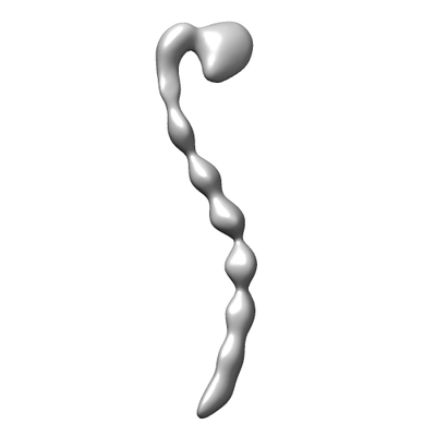 emd_8543.png emd_8543.png | 13 KB | ||
| アーカイブディレクトリ |  http://ftp.pdbj.org/pub/emdb/structures/EMD-8543ftp://ftp.pdbj.org/pub/emdb/structures/EMD-8543 http://ftp.pdbj.org/pub/emdb/structures/EMD-8543ftp://ftp.pdbj.org/pub/emdb/structures/EMD-8543 | HTTPS FTP |
-関連構造データ


-リンク
| EMDBのページ | EMDB (EBI/PDBe) / EMDataResource |
|---|
-マップ
| ファイル | ダウンロード / ファイル: emd_8543.map.gz / 形式: CCP4 / 大きさ: 22.2 MB / タイプ: IMAGE STORED AS FLOATING POINT NUMBER (4 BYTES) | ||||||||||||||||||||||||||||||||||||||||||||||||||||||||||||||||||||
|---|---|---|---|---|---|---|---|---|---|---|---|---|---|---|---|---|---|---|---|---|---|---|---|---|---|---|---|---|---|---|---|---|---|---|---|---|---|---|---|---|---|---|---|---|---|---|---|---|---|---|---|---|---|---|---|---|---|---|---|---|---|---|---|---|---|---|---|---|---|
| 注釈 | 3D negative stain EM structure of Pom152 | ||||||||||||||||||||||||||||||||||||||||||||||||||||||||||||||||||||
| ボクセルのサイズ | X=Y=Z: 2.93 Å | ||||||||||||||||||||||||||||||||||||||||||||||||||||||||||||||||||||
| 密度 |
| ||||||||||||||||||||||||||||||||||||||||||||||||||||||||||||||||||||
| 対称性 | 空間群: 1 | ||||||||||||||||||||||||||||||||||||||||||||||||||||||||||||||||||||
| 詳細 | EMDB XML:
CCP4マップ ヘッダ情報:
| ||||||||||||||||||||||||||||||||||||||||||||||||||||||||||||||||||||
-添付データ
- 試料の構成要素
-全体 : Nucleoporin Pom152
| 全体 | 名称: Nucleoporin Pom152 |
|---|---|
| 要素 |
|
-超分子 #1: Nucleoporin Pom152
| 超分子 | 名称: Nucleoporin Pom152 / タイプ: cell / ID: 1 / 親要素: 0 / 含まれる分子: all |
|---|---|
| 由来(天然) | 生物種: Saccharomyces cerevisiae (パン酵母) |
-分子 #1: Pom152
| 分子 | 名称: Pom152 / タイプ: protein_or_peptide / ID: 1 / 光学異性体: LEVO |
|---|---|
| 由来(天然) | 生物種: Saccharomyces cerevisiae (パン酵母) |
| 配列 | 文字列: MEHRYNVFND TPRGNHWMGS SVSGSPRPSY SSRPNVNTTR RFQYSDDEPA EKIRPLRSRS FKSTESNIS DEKSRISERD SKDRYINGDK KVDIYSLPLI STDVLEISKQ RTFAVILFLI I QCYKIYDL VILKSGLPLS GLLFKNYRFN FISKYFIIDS FFLYVLPSFN ...文字列: MEHRYNVFND TPRGNHWMGS SVSGSPRPSY SSRPNVNTTR RFQYSDDEPA EKIRPLRSRS FKSTESNIS DEKSRISERD SKDRYINGDK KVDIYSLPLI STDVLEISKQ RTFAVILFLI I QCYKIYDL VILKSGLPLS GLLFKNYRFN FISKYFIIDS FFLYVLPSFN IPRLTFKPWV VY LQILAML LLNIFISSDH EFVLISLIMT TWRKLYTKEL SVTGSAINHH RIFDSSAHFK GAL TIKILP ENTAMFNPLH ESYCLPMDTN LFKINSIDVP IRINSTEEIE YIELEYRDLY TNSV ELRSL SKKDFKIIDN PKSFLKKDQS VLKSHSNDFE EGSTIRYLAV TLQDIGFYQI KKIVD SKKL NLKIHQSHLV VPYCPIASIT GTGSNDRCIG DSDNVSFEIQ GVPPMKLAYS KIVNGQ TFS YVDSSLQPEY FESPLQSSKS KQSFTQGELN DLKWGRNQPV NINLDSSITQ DGKFAYK ID KITDGLGNVV DFTSLPEELK KRYDLSYNFN VHEVPRAALE ERFDPKSPTK RSIAIVFE E IKNWISDIPY VISLSYTDAQ DKSKKIMNVT TDSLTKVLQA DLPGSYNLEY IESKFCPGE IVGKSNVLVT MPVAPTMEVK SFPILDQCVG QVGLNFELSF TGAPPYYYNT KIYKLENGER KLYDAKRYT SEGTRNRFSY SPPKEGNYEI VFDTVSNKLF TEPIKLEPVK EYTFKTSMRV K PSASLKLH HDLKLCLGDH SSVPVALKGQ GPFTLTYDII ETFSSKRKTF EIKEIKTNEY VI KTPVFTT GGDYILSLVS IKDSTGCVVG LSQPDAKIQV RRDIPSAAFN FFEPIKEAKI KHG SVTEIP LKLSGEGPFT VKFKHMDYDG NIVKEFENKF QNSYKPALKV SKEGLYQLVD IRDS SCQGN VIYRNSLYKV SFLEKPKFAI QDNHHITKVT ENLFSKEEVC QGMEGTVDLA LFGSP PFIL EYDLMAPNGH ISTKKIQVAT KYASLKLPNQ IPGEYITTIK AIFDGNYGES DIHFRE HQS ELIIKQTVHP IPDVAFADGG KTLRACAANV DQISFLEPIN LKFLQGESPF SITFSVY HE STSRTDQYTI DNIDSENFSF EKLYEGMKLG NHAITIDSVV DANGCVNSLI SGPRNQIL V SITDAPKIHI LDPSTEYCVG DYVAYQLNGV APFMIKYEFN GIPLKSKERS SQFVRLASE PGIISITSLQ DSSSQCIVDF TNPKLKSEFD DLSLNIHPIP SVTVSQGNYV TEDIREGDQA EVIFSFEGT PPFSLTYVRT EETDGKHGKR RSQVVETHKV TDIYSHEYKV ITSLQGTYEA I EITDAYCF AKNDLFFNN |
-実験情報
-構造解析
| 手法 | ネガティブ染色法 |
|---|---|
 解析 解析 | 単粒子再構成法 |
| 試料の集合状態 | particle |
-試料調製
| 緩衝液 | pH: 7.4 構成要素:
詳細: Solutions were made fresh from concentrated, and sterile filtered. | |||||||||||||||||||||
|---|---|---|---|---|---|---|---|---|---|---|---|---|---|---|---|---|---|---|---|---|---|---|
| 染色 | タイプ: NEGATIVE / 材質: Uranyl formate 詳細: 3 ul of protein was added to grids. The excess sample was blotted and the grid washed several times with water. The sample was stained with several drops of uranyl formate, blotted between ...詳細: 3 ul of protein was added to grids. The excess sample was blotted and the grid washed several times with water. The sample was stained with several drops of uranyl formate, blotted between each stain. After the last staining (30 sec), the sample was blotted and air dried. | |||||||||||||||||||||
| グリッド | モデル: Electron Microscopy Sciences / 材質: COPPER / メッシュ: 200 / 支持フィルム - 材質: CARBON / 支持フィルム - トポロジー: CONTINUOUS / 前処理 - タイプ: PLASMA CLEANING / 前処理 - 雰囲気: OTHER | |||||||||||||||||||||
| 詳細 | The endogenous sample was purified using affinity tags, then natively eluted and isolated on a 5-20% sucrose gradient. Fractions identified by SDS-PAGE and mass spectrometry. |
- 電子顕微鏡法
| 顕微鏡 | JEOL 2100F |
|---|---|
| 電子線 | 加速電圧: 120 kV / 電子線源: FIELD EMISSION GUN |
| 電子光学系 | C2レンズ絞り径: 40.0 µm / 倍率(補正後): 81911 / 照射モード: FLOOD BEAM / 撮影モード: BRIGHT FIELDBright-field microscopy / Cs: 2.0 mm / 最大 デフォーカス(公称値): 1.5 µm / 最小 デフォーカス(公称値): 1.0 µm / 倍率(公称値): 50000 |
| 試料ステージ | 試料ホルダーモデル: JEOL |
| 撮影 | フィルム・検出器のモデル: TVIPS TEMCAM-F224 (2k x 2k) 撮影したグリッド数: 2 / 実像数: 1220 / 平均露光時間: 2.0 sec. / 平均電子線量: 15.0 e/Å2 詳細: Images were collected at tilt angles 0 and 40 degrees |
-画像解析
| 粒子像選択 | 選択した数: 16826 詳細: Number of particles includes 8413 untilted and 8413 tilted particles. |
|---|---|
| CTF補正 | ソフトウェア - 名称: EMAN2 (ver. Ctffind3) |
| 初期モデル | モデルのタイプ: RANDOM CONICAL TILT / Random conical tilt - Number images: 7723 / Random conical tilt - Tilt angle: 40 degrees |
| 初期 角度割当 | タイプ: OTHER / ソフトウェア - 名称: SPARX (ver. EMAN2 2.06) 詳細: Sparx was used to generate the 2D classes of the untilted particles and to get their relative orientations to derive the Euler angles, then Spider (version 17.12) was used to do the back projection. |
| 最終 3次元分類 | クラス数: 2 / ソフトウェア - 名称: RELION (ver. 1.3) |
| 最終 角度割当 | タイプ: PROJECTION MATCHING / ソフトウェア - 名称: RELION (ver. 1.3) |
| 最終 再構成 | 使用したクラス数: 1 / 想定した対称性 - 点群: C1 (非対称) / アルゴリズム: BACK PROJECTION / 解像度のタイプ: BY AUTHOR / 解像度: 25.0 Å / 解像度の算出法: FSC 0.143 CUT-OFF / ソフトウェア - 名称: RELION (ver. 1.3) / 使用した粒子像数: 7967 |
| 詳細 | The selected images were normalized. |
| FSC曲線 (解像度の算出) | 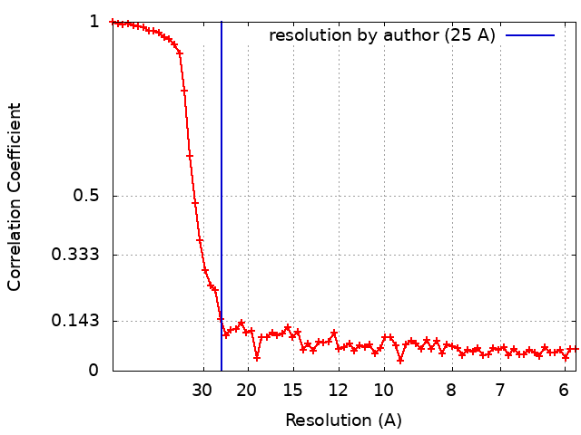 |
