Movie
Movie Controller


+ Open data

- Basic information
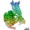
| Entry | Database: PDB / ID: 3bo0 | ||||||
|---|---|---|---|---|---|---|---|
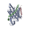
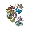
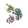
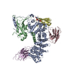
| Title | Ribosome-SecY complex | ||||||
 Components Components |
| ||||||
 Keywords Keywords | RIBOSOME / Ribosome-SecY complex / protein translocation | ||||||
| Function / homology |  Function and homology information Function and homology informationintracellular protein transmembrane transport / SRP-dependent cotranslational protein targeting to membrane, translocation / signal sequence binding / protein transmembrane transporter activity / protein secretion / protein targeting / protein transport / plasma membrane Similarity search - Function | ||||||
| Biological species |  | ||||||
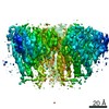
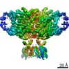
| Method | ELECTRON MICROSCOPY / single particle reconstruction / cryo EM / Resolution: 9.6 Å | ||||||
 Authors Authors | Akey, C.W. / Menetret, J.F. | ||||||
 Citation Citation | Journal: Mol Cell / Year: 2007 Title: Ribosome binding of a single copy of the SecY complex: implications for protein translocation. Authors: Jean-François Ménétret / Julia Schaletzky / William M Clemons / Andrew R Osborne / Sigrid S Skånland / Carilee Denison / Steven P Gygi / Don S Kirkpatrick / Eunyong Park / Steven J ...Authors: Jean-François Ménétret / Julia Schaletzky / William M Clemons / Andrew R Osborne / Sigrid S Skånland / Carilee Denison / Steven P Gygi / Don S Kirkpatrick / Eunyong Park / Steven J Ludtke / Tom A Rapoport / Christopher W Akey /  Abstract: The SecY complex associates with the ribosome to form a protein translocation channel in the bacterial plasma membrane. We have used cryo-electron microscopy and quantitative mass spectrometry to ...The SecY complex associates with the ribosome to form a protein translocation channel in the bacterial plasma membrane. We have used cryo-electron microscopy and quantitative mass spectrometry to show that a nontranslating E. coli ribosome binds to a single SecY complex. The crystal structure of an archaeal SecY complex was then docked into the electron density maps. In the resulting model, two cytoplasmic loops of SecY extend into the exit tunnel near proteins L23, L29, and L24. The loop between transmembrane helices 8 and 9 interacts with helices H59 and H50 in the large subunit RNA, while the 6/7 loop interacts with H7. We also show that point mutations of basic residues within either loop abolish ribosome binding. We suggest that SecY binds to this primary site on the ribosome and subsequently captures and translocates the nascent chain. | ||||||
| History |
|
- Structure visualization
| Movie |
Movie viewer |
|---|---|
| Structure viewer | Molecule: MolmilJmol/JSmol |
- Downloads & links
-Download
| PDBx/mmCIF format | 3bo0.cif.gz | 161.9 KB | Display | PDBx/mmCIF format |
|---|---|---|---|---|
| PDB format | pdb3bo0.ent.gz | 122.6 KB | Display | PDB format |
| PDBx/mmJSON format | 3bo0.json.gz | Tree view | PDBx/mmJSON format | |
| Others |  Other downloads Other downloads |
-Validation report
| Summary document | 3bo0_validation.pdf.gz | 809.4 KB | Display | wwPDB validaton report |
|---|---|---|---|---|
| Full document | 3bo0_full_validation.pdf.gz | 911.4 KB | Display | |
| Data in XML | 3bo0_validation.xml.gz | 37.5 KB | Display | |
| Data in CIF | 3bo0_validation.cif.gz | 54.5 KB | Display | |
| Arichive directory | https://data.pdbj.org/pub/pdb/validation_reports/bo/3bo0ftp://data.pdbj.org/pub/pdb/validation_reports/bo/3bo0 | HTTPS FTP |
-Related structure data
| Related structure data | 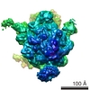 1484MC 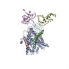 3bo1C C: citing same article ( M: map data used to model this data |
|---|---|
| Similar structure data |

-Links
 PDBj
PDBj


- Assembly
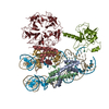
| Deposited unit | 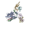
|
|---|---|
| 1 |
|
-Components
-23S RIBOSOMAL ... , 4 types, 4 molecules DEFG
| #1: RNA chain | Mass: 8698.224 Da / Num. of mol.: 1 / Fragment: GB residues 79-105 / Source method: isolated from a natural source / Details: helix 7 / Source: (natural) |
|---|---|
| #2: RNA chain | Mass: 8860.439 Da / Num. of mol.: 1 / Fragment: GB residues 478-504 / Source method: isolated from a natural source / Details: helix 24 / Source: (natural) |
| #3: RNA chain | Mass: 6024.601 Da / Num. of mol.: 1 / Fragment: GB residues 1385-1403 / Source method: isolated from a natural source / Details: helix 47 / Source: (natural) |
| #4: RNA chain | Mass: 10382.260 Da / Num. of mol.: 1 / Fragment: GB residues 1518-1549 / Source method: isolated from a natural source / Details: helix 59 / Source: (natural) |
-PREPROTEIN TRANSLOCASE ... , 3 types, 3 molecules ABC
| #5: Protein | Mass: 48367.695 Da / Num. of mol.: 1 Source method: isolated from a genetically manipulated source Source: (gene. exp.) |
|---|---|
| #6: Protein | Mass: 7149.573 Da / Num. of mol.: 1 Source method: isolated from a genetically manipulated source Source: (gene. exp.) |
| #7: Protein/peptide | Mass: 3639.310 Da / Num. of mol.: 1 Source method: isolated from a genetically manipulated source Source: (gene. exp.) |
-Details
| Sequence details | THIS PDB FILE WAS OBTAINED UPON RIGID BODY REFINEMENT OF A STARTING PDB FILE INTO A CRYO-EM MAP. ...THIS PDB FILE WAS OBTAINED UPON RIGID BODY REFINEMENT |
|---|
-Experimental details
-Experiment
| Experiment | Method: ELECTRON MICROSCOPY |
|---|---|
| EM experiment | Aggregation state: PARTICLE / 3D reconstruction method: single particle reconstruction |
- Sample preparation
| Component | Name: Ribosome-SecY complex / Type: RIBOSOME / Details: in DDM |
|---|---|
| Buffer solution | Name: 50 mM HEPES- KOH pH 7.5, 100 mM KOAc, 10 mM Mg(OAc)2, 0.05% DDM pH: 7.5 Details: 50 mM HEPES- KOH pH 7.5, 100 mM KOAc, 10 mM Mg(OAc)2, 0.05% DDM |
| Specimen | Embedding applied: NO / Shadowing applied: NO / Staining applied: NO / Vitrification applied: YES |
| Specimen support | Details: Solid carbon on a holey film, 400 mesh Cu grid |
| Vitrification | Instrument: HOMEMADE PLUNGER / Cryogen name: ETHANE Details: The specimens were plunge frozen in liquid ethane at 4 degrees C at an RH of ~90-95%. |
- Electron microscopy imaging
| Experimental equipment |  Model: Tecnai F20 / Image courtesy: FEI Company |
|---|---|
| Microscopy | Model: FEI TECNAI F20 / Details: Gatan DH626 cold holder |
| Electron gun | Electron source:  FIELD EMISSION GUN / Accelerating voltage: 200 kV / Illumination mode: FLOOD BEAM FIELD EMISSION GUN / Accelerating voltage: 200 kV / Illumination mode: FLOOD BEAM |
| Electron lens | Mode: BRIGHT FIELD / Nominal magnification: 50000 X / Calibrated magnification: 51000 X / Nominal defocus max: -3000 nm / Nominal defocus min: -700 nm / Cs: 2 mm |
| Specimen holder | Temperature: 93 K / Tilt angle max: 0 ° / Tilt angle min: 0 ° |
| Image recording | Electron dose: 15 e/Å2 / Film or detector model: KODAK SO-163 FILM |
- Processing
| CTF correction | Details: EMAN- phase flipping of particles form the same micrograph | |||||||||||||||||||||
|---|---|---|---|---|---|---|---|---|---|---|---|---|---|---|---|---|---|---|---|---|---|---|
| Symmetry | Point symmetry: C1 (asymmetric) | |||||||||||||||||||||
| 3D reconstruction | Method: Projection matching in EMAN / Resolution: 9.6 Å / Num. of particles: 39000 / Actual pixel size: 2.73 Å / Symmetry type: POINT | |||||||||||||||||||||
| Atomic model building | Protocol: RIGID BODY FIT / Space: REAL / Target criteria: visual fit in O and Chimera Details: REFINEMENT PROTOCOL--rigid body, followed by manual rebuilding and extension of the loops | |||||||||||||||||||||
| Atomic model building |
| |||||||||||||||||||||
| Refinement step | Cycle: LAST
|