- PDB-2mme: Hybrid structure of the Shigella flexneri MxiH Type three secreti... -
+
Open data
ID or keywords:
Loading...
-
Basic information
Entry
Database: PDB / ID: 2mme
Title
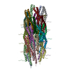
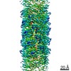
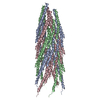
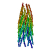
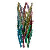
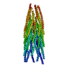
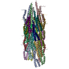
Hybrid structure of the Shigella flexneri MxiH Type three secretion system needle
Components
MxiH
Keywords
PROTEIN TRANSPORT / type-three secretion system / filamentous protein / helical assembly / Shigella flexneri / protein translocation / hybrid methods / Rosetta
Function / homology
Type III secretion, needle-protein-like / Type III secretion, needle-protein-like superfamily / Type III secretion needle MxiH, YscF, SsaG, EprI, PscF, EscF / Type III secretion system, needle protein / type III protein secretion system complex / protein secretion by the type III secretion system / : / MxiH
Function and homology information
Biological species
Shigella flexneri (bacteria)
Method
SOLID-STATE NMR / ELECTRON MICROSCOPY / helical reconstruction / Rosetta fold-and-dock, Rosetta symmetric relax / cryo EM / Resolution: 7.7 Å
Journal: Proc Natl Acad Sci U S A / Year: 2012 Title: Structure of a type III secretion needle at 7-Å resolution provides insights into its assembly and signaling mechanisms. Authors: Takashi Fujii / Martin Cheung / Amandine Blanco / Takayuki Kato / Ariel J Blocker / Keiichi Namba / Abstract: Type III secretion systems of Gram-negative bacteria form injection devices that deliver effector proteins into eukaryotic cells during infection. They span both bacterial membranes and the ...Type III secretion systems of Gram-negative bacteria form injection devices that deliver effector proteins into eukaryotic cells during infection. They span both bacterial membranes and the extracellular space to connect with the host cell plasma membrane. Their extracellular portion is a needle-like, hollow tube that serves as a secretion conduit for effector proteins. The needle of Shigella flexneri is approximately 50-nm long and 7-nm thick and is made by the helical assembly of one protein, MxiH. We provide a 7-Å resolution 3D image reconstruction of the Shigella needle by electron cryomicroscopy, which resolves α-helices and a β-hairpin that has never been observed in the crystal and solution structures of needle proteins, including MxiH. An atomic model of the needle based on the 3D-density map, in comparison with that of the bacterial-flagellar filament, provides insights into how such a thin tubular structure is stably assembled by intricate intermolecular interactions. The map also illuminates how the needle-length control protein functions as a ruler within the central channel during export of MxiH for assembly at the distal end of the needle, and how the secretion-activation signal may be transduced through a conformational change of the needle upon host-cell contact.
THIS ENTRY 2MME CONTAINS A STRUCTURAL MODEL FIT TO AN ELECTRON MICROSCOPY MAP (EMD-5352) DETERMINED ...THIS ENTRY 2MME CONTAINS A STRUCTURAL MODEL FIT TO AN ELECTRON MICROSCOPY MAP (EMD-5352) DETERMINED ORIGINALLY BY AUTHORS: T.FUJII, M.CHEUNG, A.BLANCO, T.KATO, A.J.BLOCKER, K.NAMBA
Electron source: FIELD EMISSION GUN / Accelerating voltage: 200 kV / Illumination mode: FLOOD BEAM
Electron lens
Mode: BRIGHT FIELD / Nominal magnification: 50000 X / Nominal defocus max: 2000 nm / Nominal defocus min: 1000 nm
Image recording
Electron dose: 20 e/Å2 / Film or detector model: TVIPS TEMCAM-F415 (4k x 4k)
NMR spectrometer
Type: Bruker Avance / Manufacturer: Bruker / Model: AVANCE / Field strength: 850 MHz
-
Processing
3D reconstruction
Resolution: 7.7 Å / Symmetry type: HELICAL
Refinement step
Cycle: LAST
Protein
Nucleic acid
Ligand
Solvent
Total
Num. atoms
18763
0
0
0
18763
NMR software
Name
Developer
Classification
Sparky
Goddard
dataanalysis
Rosetta
refinement
Refinement
Method: Rosetta fold-and-dock, Rosetta symmetric relax / Software ordinal: 2 Details: symmetric fragment-based Monte Carlo trials followed by full-atom refinement, symmetric refinement (relax) of backbone, sidechain, and rigid-body degrees of freedom
NMR representative
Selection criteria: lowest energy
NMR ensemble
Conformer selection criteria: target function / Conformers calculated total number: 5000 / Conformers submitted total number: 10
+
About Yorodumi
-
News
-
Feb 9, 2022. New format data for meta-information of EMDB entries
New format data for meta-information of EMDB entries
Version 3 of the EMDB header file is now the official format.
The previous official version 1.9 will be removed from the archive.
In the structure databanks used in Yorodumi, some data are registered as the other names, "COVID-19 virus" and "2019-nCoV". Here are the details of the virus and the list of structure data.
Jan 31, 2019. EMDB accession codes are about to change! (news from PDBe EMDB page)
EMDB accession codes are about to change! (news from PDBe EMDB page)
The allocation of 4 digits for EMDB accession codes will soon come to an end. Whilst these codes will remain in use, new EMDB accession codes will include an additional digit and will expand incrementally as the available range of codes is exhausted. The current 4-digit format prefixed with “EMD-” (i.e. EMD-XXXX) will advance to a 5-digit format (i.e. EMD-XXXXX), and so on. It is currently estimated that the 4-digit codes will be depleted around Spring 2019, at which point the 5-digit format will come into force.
The EM Navigator/Yorodumi systems omit the EMD- prefix.
Related info.:Q: What is EMD? / ID/Accession-code notation in Yorodumi/EM Navigator
Yorodumi is a browser for structure data from EMDB, PDB, SASBDB, etc.
This page is also the successor to EM Navigator detail page, and also detail information page/front-end page for Omokage search.
The word "yorodu" (or yorozu) is an old Japanese word meaning "ten thousand". "mi" (miru) is to see.
Related info.:EMDB / PDB / SASBDB / Comparison of 3 databanks / Yorodumi Search / Aug 31, 2016. New EM Navigator & Yorodumi / Yorodumi Papers / Jmol/JSmol / Function and homology information / Changes in new EM Navigator and Yorodumi
 Movie
Movie Controller

 Yorodumi
 Open data
 Basic information
 Components
 Keywords
 Function and homology information
 Shigella flexneri (bacteria)
 Authors
 Citation
 Structure visualization
 Downloads & links
 Other downloads
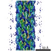

 PDBj
PDBj Assembly
 Sample preparation
 FIELD EMISSION GUN / Accelerating voltage: 200 kV / Illumination mode: FLOOD BEAM
 Processing