ムービー
ムービー コントローラー


+ データを開く

- 基本情報
基本情報
| 登録情報 | データベース: EMDB / ID: EMD-9104 | |||||||||
|---|---|---|---|---|---|---|---|---|---|---|
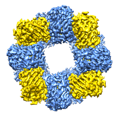
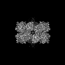
| タイトル | Cryo-EM structure of the Ceru+32/GFP-17 protomer | |||||||||
 マップデータ マップデータ | The structure of a highly symmetrical, well-defined 16-mer formed by 8 Ceru+32 and 8 GFP-17 | |||||||||
 試料 試料 |
| |||||||||
 キーワード キーワード | Supercharged protein assembly / electrostatic interactions / 16-mer / D4 symmetry / LUMINESCENT PROTEIN | |||||||||
| 機能・相同性 | Green fluorescent protein, GFP / Green fluorescent protein-related / Green fluorescent protein / Green fluorescent protein / bioluminescence / generation of precursor metabolites and energy / Green fluorescent protein 機能・相同性情報 機能・相同性情報 | |||||||||
| 生物種 |   Aequorea victoria (オワンクラゲ) Aequorea victoria (オワンクラゲ) | |||||||||
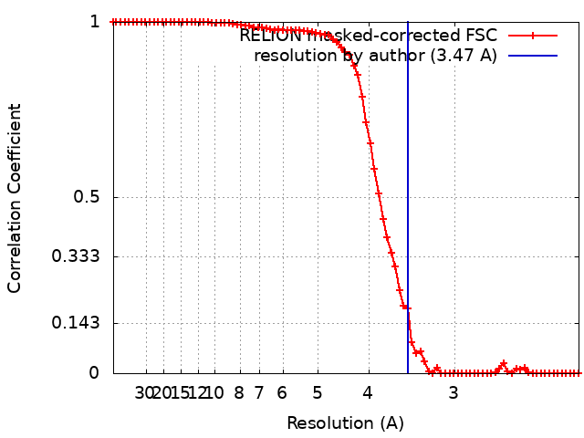
| 手法 | 単粒子再構成法 / クライオ電子顕微鏡法 / 解像度: 3.47 Å | |||||||||
 データ登録者 データ登録者 | Simon AJ / Zhou Y | |||||||||
| 資金援助 |  米国, 1件 米国, 1件
| |||||||||
 引用 引用 | ジャーナル: Nat Chem / 年: 2019 タイトル: Supercharging enables organized assembly of synthetic biomolecules. 著者: Anna J Simon / Yi Zhou / Vyas Ramasubramani / Jens Glaser / Arti Pothukuchy / Jimmy Gollihar / Jillian C Gerberich / Janelle C Leggere / Barrett R Morrow / Cheulhee Jung / Sharon C Glotzer / ...著者: Anna J Simon / Yi Zhou / Vyas Ramasubramani / Jens Glaser / Arti Pothukuchy / Jimmy Gollihar / Jillian C Gerberich / Janelle C Leggere / Barrett R Morrow / Cheulhee Jung / Sharon C Glotzer / David W Taylor / Andrew D Ellington /  要旨: Symmetrical protein oligomers are ubiquitous in biological systems and perform key structural and regulatory functions. However, there are few methods for constructing such oligomers. Here we have ...Symmetrical protein oligomers are ubiquitous in biological systems and perform key structural and regulatory functions. However, there are few methods for constructing such oligomers. Here we have engineered completely synthetic, symmetrical oligomers by combining pairs of oppositely supercharged variants of a normally monomeric model protein through a strategy we term 'supercharged protein assembly' (SuPrA). We show that supercharged variants of green fluorescent protein can assemble into a variety of architectures including a well-defined symmetrical 16-mer structure that we solved using cryo-electron microscopy at 3.47 Å resolution. The 16-mer is composed of two stacked rings of octamers, in which the octamers contain supercharged proteins of alternating charges, and interactions within and between the rings are mediated by a variety of specific electrostatic contacts. The ready assembly of this structure suggests that combining oppositely supercharged pairs of protein variants may provide broad opportunities for generating novel architectures via otherwise unprogrammed interactions. | |||||||||
| 履歴 |
|
- 構造の表示
構造の表示
| ムービー |
ムービービューア |
|---|---|
| 構造ビューア | EMマップ: SurfViewMolmilJmol/JSmol |
| 添付画像 |
- ダウンロードとリンク
-EMDBアーカイブ
| マップデータ | emd_9104.map.gz | 40.1 MB | EMDBマップデータ形式 | |
|---|---|---|---|---|
| ヘッダ (付随情報) | emd-9104-v30.xmlemd-9104.xml | 13.4 KB 13.4 KB | 表示 表示 | EMDBヘッダ |
| FSC (解像度算出) | emd_9104_fsc.xml | 7.8 KB | 表示 | FSCデータファイル |
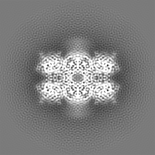
| 画像 |  emd_9104.png emd_9104.png | 223.5 KB | ||
| Filedesc metadata | emd-9104.cif.gz | 6 KB | ||
| アーカイブディレクトリ |  http://ftp.pdbj.org/pub/emdb/structures/EMD-9104ftp://ftp.pdbj.org/pub/emdb/structures/EMD-9104 http://ftp.pdbj.org/pub/emdb/structures/EMD-9104ftp://ftp.pdbj.org/pub/emdb/structures/EMD-9104 | HTTPS FTP |
-関連構造データ


-リンク
| EMDBのページ | EMDB (EBI/PDBe) / EMDataResource |
|---|---|
| 「今月の分子」の関連する項目 |


-マップ
| ファイル | ダウンロード / ファイル: emd_9104.map.gz / 形式: CCP4 / 大きさ: 42.9 MB / タイプ: IMAGE STORED AS FLOATING POINT NUMBER (4 BYTES) | ||||||||||||||||||||||||||||||||||||||||||||||||||||||||||||
|---|---|---|---|---|---|---|---|---|---|---|---|---|---|---|---|---|---|---|---|---|---|---|---|---|---|---|---|---|---|---|---|---|---|---|---|---|---|---|---|---|---|---|---|---|---|---|---|---|---|---|---|---|---|---|---|---|---|---|---|---|---|
| 注釈 | The structure of a highly symmetrical, well-defined 16-mer formed by 8 Ceru+32 and 8 GFP-17 | ||||||||||||||||||||||||||||||||||||||||||||||||||||||||||||
| 投影像・断面図 | 画像のコントロール
画像は Spider により作成 | ||||||||||||||||||||||||||||||||||||||||||||||||||||||||||||
| ボクセルのサイズ | X=Y=Z: 1.1 Å | ||||||||||||||||||||||||||||||||||||||||||||||||||||||||||||
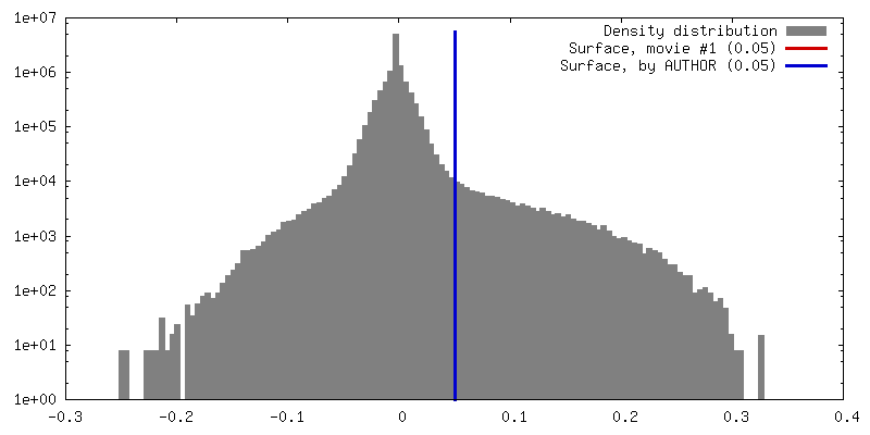
| 密度 |
| ||||||||||||||||||||||||||||||||||||||||||||||||||||||||||||
| 対称性 | 空間群: 1 | ||||||||||||||||||||||||||||||||||||||||||||||||||||||||||||
| 詳細 | EMDB XML:
CCP4マップ ヘッダ情報:
| ||||||||||||||||||||||||||||||||||||||||||||||||||||||||||||
 Z (Sec.)
 Y (Row.)
 X (Col.)


-添付データ
- 試料の構成要素
-全体 : Ceru+32/GFP-17 protomer
| 全体 | 名称: Ceru+32/GFP-17 protomer |
|---|---|
| 要素 |
|
-超分子 #1: Ceru+32/GFP-17 protomer
| 超分子 | 名称: Ceru+32/GFP-17 protomer / タイプ: complex / ID: 1 / 親要素: 0 / 含まれる分子: all |
|---|---|
| 由来(天然) | 生物種: Aequorea victoria (オワンクラゲ) |
| 分子量 | 理論値: 430 KDa |
-分子 #1: Ceru+32
| 分子 | 名称: Ceru+32 / タイプ: protein_or_peptide / ID: 1 詳細: Mutations present in the construct but not listed in the mutation list are derived from the reference construct (PDB entry 2B3P). コピー数: 8 / 光学異性体: LEVO |
|---|---|
| 由来(天然) | 生物種: Aequorea victoria (オワンクラゲ) |
| 分子量 | 理論値: 28.425195 KDa |
| 組換発現 | 生物種:  |
| 配列 | 文字列: MASKGERLFR GKVPILVELK GDVNGHKFSV RGKGKGDATN GKLTLKFICT TGKLPVPWPT LVTTLTWGVQ CFARYPKHMK RHDFFKSAM PKGYVQERTI SFKKDGTYKT RAEVKFEGRT LVNRIKLKGR DFKEKGNILG HKLRYNGISD KVYITADKRK N GIKAKFKI ...文字列: MASKGERLFR GKVPILVELK GDVNGHKFSV RGKGKGDATN GKLTLKFICT TGKLPVPWPT LVTTLTWGVQ CFARYPKHMK RHDFFKSAM PKGYVQERTI SFKKDGTYKT RAEVKFEGRT LVNRIKLKGR DFKEKGNILG HKLRYNGISD KVYITADKRK N GIKAKFKI RHNVKDGSVQ LADHYQQNTP IGRGPVLLPR NHYLSTRSVL SKDPKEKRDH MVLLEFVTAA GIKHGRDERY KL EHHHHHH UniProtKB: Green fluorescent protein |
-分子 #2: GFP-17
| 分子 | 名称: GFP-17 / タイプ: protein_or_peptide / ID: 2 詳細: Mutations present in the construct but not listed in the mutation list are derived from the reference construct (PDB entry 2B3P). コピー数: 8 / 光学異性体: LEVO |
|---|---|
| 由来(天然) | 生物種: Aequorea victoria (オワンクラゲ) |
| 分子量 | 理論値: 27.226242 KDa |
| 組換発現 | 生物種: |
| 配列 | 文字列: MSKGEELFTG VVPILVELDG DVNGHKFSVR GEGEGDADNG KLDLKFICTT GKLPVPWPTL VTTLTYGVQC FSRYPDHMKE HDFFKSAMP EGYVQERTIS FKDDGTYKTR AEVKFEGDTL VNRIELKGID FKEDGNILGH KLEYNFNSHE VYITADDEKN G IKAEFKIR ...文字列: MSKGEELFTG VVPILVELDG DVNGHKFSVR GEGEGDADNG KLDLKFICTT GKLPVPWPTL VTTLTYGVQC FSRYPDHMKE HDFFKSAMP EGYVQERTIS FKDDGTYKTR AEVKFEGDTL VNRIELKGID FKEDGNILGH KLEYNFNSHE VYITADDEKN G IKAEFKIR HNVEDGSVQL ADHYQQNTPI GDGPDLLPDE HYLSTQSVLS KDPNEKRDHM VLLEFVTADG ITEGHHHHHH HH UniProtKB: Green fluorescent protein |
-実験情報
-構造解析
| 手法 | クライオ電子顕微鏡法 |
|---|---|
 解析 解析 | 単粒子再構成法 |
| 試料の集合状態 | particle |
-試料調製
| 緩衝液 | pH: 7.4 |
|---|---|
| グリッド | モデル: C-flat-1.2/1.3 4C / 材質: COPPER / 支持フィルム - 材質: CARBON / 支持フィルム - トポロジー: HOLEY / 前処理 - タイプ: PLASMA CLEANING / 前処理 - 時間: 30 sec. |
| 凍結 | 凍結剤: ETHANE / チャンバー内湿度: 100 % / 装置: FEI VITROBOT MARK IV |
- 電子顕微鏡法
| 顕微鏡 | FEI TITAN KRIOS |
|---|---|
| 撮影 | フィルム・検出器のモデル: GATAN K2 SUMMIT (4k x 4k) 検出モード: COUNTING / 平均露光時間: 6.0 sec. / 平均電子線量: 40.0 e/Å2 |
| 電子線 | 加速電圧: 300 kV / 電子線源:  FIELD EMISSION GUN FIELD EMISSION GUN |
| 電子光学系 | C2レンズ絞り径: 100.0 µm / 照射モード: FLOOD BEAM / 撮影モード: BRIGHT FIELD / 最大 デフォーカス(公称値): 3.0 µm / 最小 デフォーカス(公称値): 1.5 µm / 倍率(公称値): 22500 |
| 試料ステージ | 試料ホルダーモデル: FEI TITAN KRIOS AUTOGRID HOLDER ホルダー冷却材: NITROGEN |
| 実験機器 |  モデル: Titan Krios / 画像提供: FEI Company |