 Movie
Movie Controller


+ Open data

- Basic information
| Entry | Database: PDB / ID: 6mdr | ||||||
|---|---|---|---|---|---|---|---|
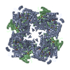
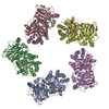
| Title | Cryo-EM structure of the Ceru+32/GFP-17 protomer | ||||||
 Components Components |
| ||||||
 Keywords Keywords | LUMINESCENT PROTEIN / Supercharged protein assembly / electrostatic interactions / 16-mer / D4 symmetry | ||||||
| Function / homology |  Function and homology information Function and homology information | ||||||
| Biological species |   Aequorea victoria (jellyfish) Aequorea victoria (jellyfish) | ||||||
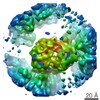
| Method | ELECTRON MICROSCOPY / single particle reconstruction / cryo EM / Resolution: 3.47 Å | ||||||
 Authors Authors | Simon, A.J. / Zhou, Y. / Ramasubramani, V. / Glaser, J. / Pothukuchy, A. / Golihar, J. / Gerberich, J.C. / Leggere, J.C. / Morrow, B.R. / Jung, C. ...Simon, A.J. / Zhou, Y. / Ramasubramani, V. / Glaser, J. / Pothukuchy, A. / Golihar, J. / Gerberich, J.C. / Leggere, J.C. / Morrow, B.R. / Jung, C. / Glotzer, S.C. / Taylor, D.W. / Ellington, A.D. | ||||||
| Funding support |  United States, 1items United States, 1items
| ||||||
 Citation Citation | Journal: Nat Chem / Year: 2019 Title: Supercharging enables organized assembly of synthetic biomolecules. Authors: Anna J Simon / Yi Zhou / Vyas Ramasubramani / Jens Glaser / Arti Pothukuchy / Jimmy Gollihar / Jillian C Gerberich / Janelle C Leggere / Barrett R Morrow / Cheulhee Jung / Sharon C Glotzer / ...Authors: Anna J Simon / Yi Zhou / Vyas Ramasubramani / Jens Glaser / Arti Pothukuchy / Jimmy Gollihar / Jillian C Gerberich / Janelle C Leggere / Barrett R Morrow / Cheulhee Jung / Sharon C Glotzer / David W Taylor / Andrew D Ellington /  Abstract: Symmetrical protein oligomers are ubiquitous in biological systems and perform key structural and regulatory functions. However, there are few methods for constructing such oligomers. Here we have ...Symmetrical protein oligomers are ubiquitous in biological systems and perform key structural and regulatory functions. However, there are few methods for constructing such oligomers. Here we have engineered completely synthetic, symmetrical oligomers by combining pairs of oppositely supercharged variants of a normally monomeric model protein through a strategy we term 'supercharged protein assembly' (SuPrA). We show that supercharged variants of green fluorescent protein can assemble into a variety of architectures including a well-defined symmetrical 16-mer structure that we solved using cryo-electron microscopy at 3.47 Å resolution. The 16-mer is composed of two stacked rings of octamers, in which the octamers contain supercharged proteins of alternating charges, and interactions within and between the rings are mediated by a variety of specific electrostatic contacts. The ready assembly of this structure suggests that combining oppositely supercharged pairs of protein variants may provide broad opportunities for generating novel architectures via otherwise unprogrammed interactions. | ||||||
| History |
|
- Structure visualization
| Movie |
Movie viewer |
|---|---|
| Structure viewer | Molecule: MolmilJmol/JSmol |
- Downloads & links
-Download
| PDBx/mmCIF format | 6mdr.cif.gz | 633.7 KB | Display | PDBx/mmCIF format |
|---|---|---|---|---|
| PDB format | pdb6mdr.ent.gz | 529.2 KB | Display | PDB format |
| PDBx/mmJSON format | 6mdr.json.gz | Tree view | PDBx/mmJSON format | |
| Others |  Other downloads Other downloads |
-Validation report
| Arichive directory | https://data.pdbj.org/pub/pdb/validation_reports/md/6mdrftp://data.pdbj.org/pub/pdb/validation_reports/md/6mdr | HTTPS FTP |
|---|
-Related structure data
| Related structure data |  9104MC M: map data used to model this data C: citing same article ( |
|---|---|
| Similar structure data |

-Links
 PDBj
PDBj

- Assembly
| Deposited unit | 
|
|---|---|
| 1 |
|
-Components
| #1: Protein | Mass: 28425.195 Da / Num. of mol.: 8 / Fragment: UNP residues 3-232 Mutation: E7R, T10R, V12K, D20K, E33K, E35K, Y67W, S73A, D77K, E91K, D103K, D118R, E125K, I129R, D134K, E143R, F146G, N147I, H149D, N150K, Q158R, N165K, E173K, D191R, D198R, Q205R, N213K, T231K Source method: isolated from a genetically manipulated source Details: Mutations present in the construct but not listed in the mutation list are derived from the reference construct (PDB entry 2B3P). Source: (gene. exp.) Aequorea victoria (jellyfish) / Gene: GFP / Plasmid: pET21 / Production host:  #2: Protein | Mass: 27226.242 Da / Num. of mol.: 8 / Fragment: UNP residues 3-232 Mutation: T39D, T44D, R81E, N150E, K157D, Q158E, N165E, V194D, N199E, A228D, H232E Source method: isolated from a genetically manipulated source Details: Mutations present in the construct but not listed in the mutation list are derived from the reference construct (PDB entry 2B3P). Source: (gene. exp.) Aequorea victoria (jellyfish) / Gene: GFP / Plasmid: pET21 / Production host: Sequence details | Mutations present in the construct but not listed in the mutation list are derived from the ...Mutations present in the construct but not listed in the mutation list are derived from the reference construct (PDB entry 2B3P). | |
|---|
-Experimental details
-Experiment
| Experiment | Method: ELECTRON MICROSCOPY |
|---|---|
| EM experiment | Aggregation state: PARTICLE / 3D reconstruction method: single particle reconstruction |
- Sample preparation
| Component | Name: Ceru+32/GFP-17 protomer / Type: COMPLEX / Entity ID: all / Source: RECOMBINANT |
|---|---|
| Molecular weight | Value: 0.430 MDa / Experimental value: NO |
| Source (natural) | Organism: Aequorea victoria (jellyfish) |
| Source (recombinant) | Organism: |
| Buffer solution | pH: 7.4 |
| Specimen | Embedding applied: NO / Shadowing applied: NO / Staining applied: NO / Vitrification applied: YES |
| Specimen support | Grid material: COPPER / Grid type: C-flat-1.2/1.3 4C |
| Vitrification | Instrument: FEI VITROBOT MARK IV / Cryogen name: ETHANE / Humidity: 100 % |
- Electron microscopy imaging
| Experimental equipment |  Model: Titan Krios / Image courtesy: FEI Company |
|---|---|
| Microscopy | Model: FEI TITAN KRIOS |
| Electron gun | Electron source:  FIELD EMISSION GUN / Accelerating voltage: 300 kV / Illumination mode: FLOOD BEAM FIELD EMISSION GUN / Accelerating voltage: 300 kV / Illumination mode: FLOOD BEAM |
| Electron lens | Mode: BRIGHT FIELD / Nominal magnification: 22500 X / Nominal defocus max: 3000 nm / Nominal defocus min: 1500 nm / C2 aperture diameter: 100 µm / Alignment procedure: COMA FREE |
| Specimen holder | Cryogen: NITROGEN / Specimen holder model: FEI TITAN KRIOS AUTOGRID HOLDER |
| Image recording | Average exposure time: 6 sec. / Electron dose: 40 e/Å2 / Detector mode: COUNTING / Film or detector model: GATAN K2 SUMMIT (4k x 4k) |
| Image scans | Movie frames/image: 20 |
- Processing
| EM software |
| ||||||||||||||||||
|---|---|---|---|---|---|---|---|---|---|---|---|---|---|---|---|---|---|---|---|
| CTF correction | Type: PHASE FLIPPING ONLY | ||||||||||||||||||
| 3D reconstruction | Resolution: 3.47 Å / Resolution method: FSC 0.143 CUT-OFF / Num. of particles: 126822 / Symmetry type: POINT | ||||||||||||||||||
| Atomic model building | B value: 196 / Protocol: RIGID BODY FIT / Space: REAL | ||||||||||||||||||
| Atomic model building | PDB-ID: 2B3P Accession code: 2B3P / Source name: PDB / Type: experimental model |

