ムービー
ムービー コントローラー


+ データを開く

- 基本情報
基本情報
| 登録情報 | データベース: EMDB / ID: EMD-8912 | |||||||||
|---|---|---|---|---|---|---|---|---|---|---|
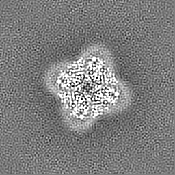
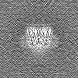
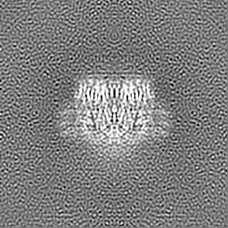
| タイトル | Human Polycsytin 2-l1 | |||||||||
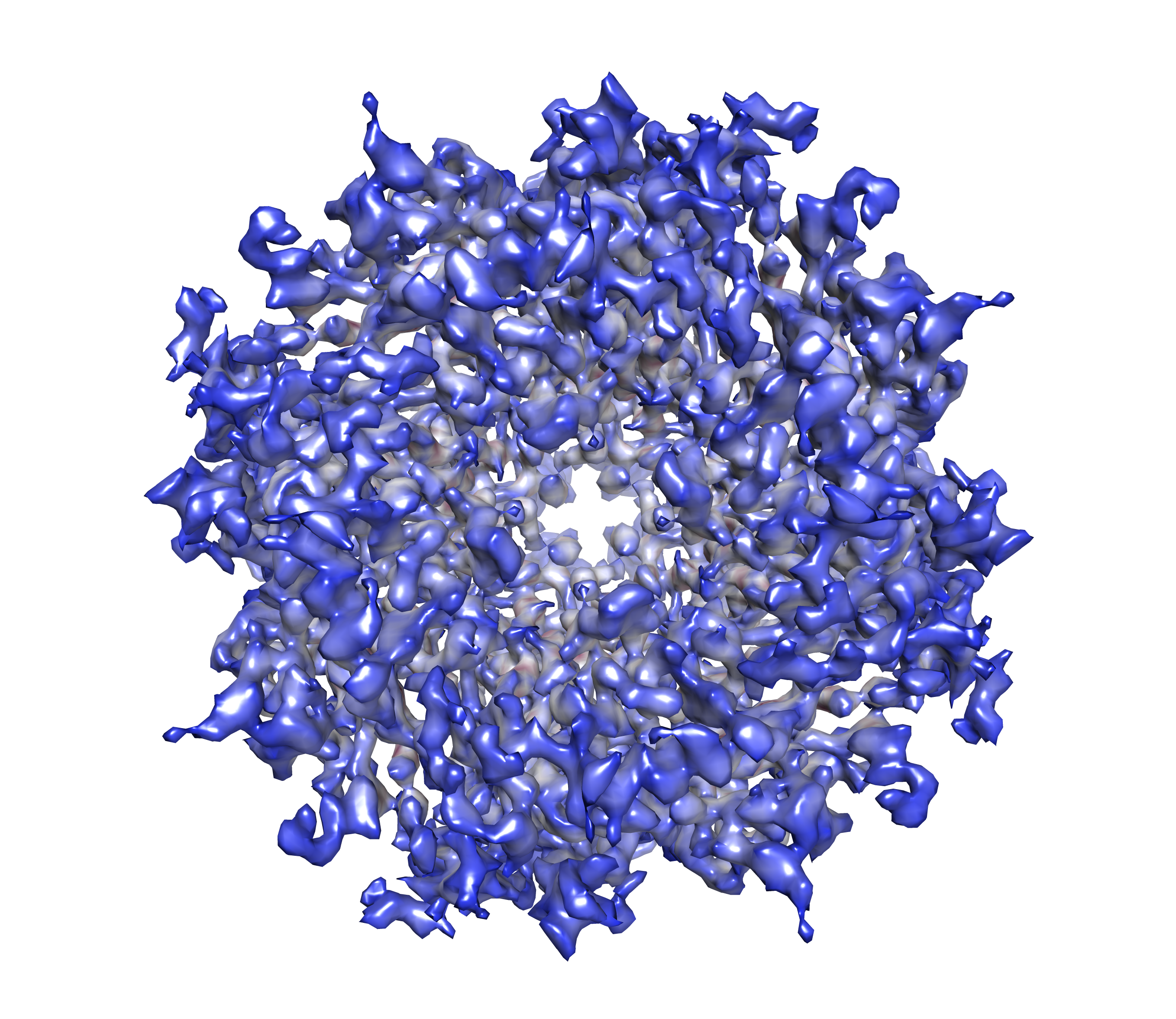
 マップデータ マップデータ | Human Polycsytin 2-l1, primary map | |||||||||
 試料 試料 |
| |||||||||
 キーワード キーワード | polycystic 2-l1 / pc2l1 / pkd2l1 / TRPP2 / MEMBRANE PROTEIN | |||||||||
| 機能・相同性 |  機能・相同性情報 機能・相同性情報sour taste receptor activity / detection of chemical stimulus involved in sensory perception of sour taste / detection of chemical stimulus involved in sensory perception of taste / sensory perception of sour taste / response to water / osmolarity-sensing monoatomic cation channel activity / pH-gated monoatomic ion channel activity / calcium-activated potassium channel activity / muscle alpha-actinin binding / detection of mechanical stimulus ...sour taste receptor activity / detection of chemical stimulus involved in sensory perception of sour taste / detection of chemical stimulus involved in sensory perception of taste / sensory perception of sour taste / response to water / osmolarity-sensing monoatomic cation channel activity / pH-gated monoatomic ion channel activity / calcium-activated potassium channel activity / muscle alpha-actinin binding / detection of mechanical stimulus / calcium-activated cation channel activity / non-motile cilium / : / ciliary membrane / smoothened signaling pathway / sodium channel activity / alpha-actinin binding / monoatomic cation transmembrane transport / monoatomic cation transport / monoatomic cation channel activity / cellular response to acidic pH / cytoskeletal protein binding / potassium ion transmembrane transport / calcium channel complex / sodium ion transmembrane transport / calcium channel activity / actin cytoskeleton / cytoplasmic vesicle / protein homotetramerization / transmembrane transporter binding / signaling receptor complex / calcium ion binding / cell surface / endoplasmic reticulum / membrane / identical protein binding / plasma membrane / cytosol 類似検索 - 分子機能 | |||||||||
| 生物種 |  Homo sapiens (ヒト) Homo sapiens (ヒト) | |||||||||
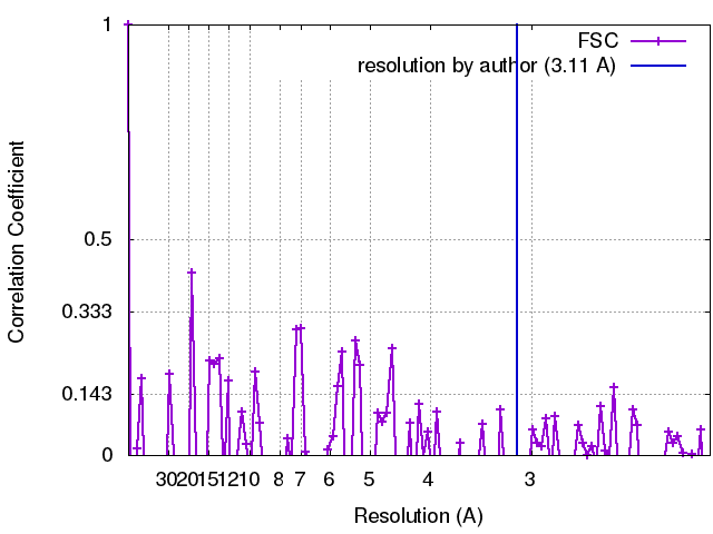
| 手法 | 単粒子再構成法 / クライオ電子顕微鏡法 / 解像度: 3.11 Å | |||||||||
 データ登録者 データ登録者 | Hulse RE / Clapham DE | |||||||||
| 資金援助 |  米国, 1件 米国, 1件
| |||||||||
 引用 引用 | ジャーナル: Elife / 年: 2018 タイトル: Cryo-EM structure of the polycystin 2-l1 ion channel. 著者: Raymond E Hulse / Zongli Li / Rick K Huang / Jin Zhang / David E Clapham /  要旨: We report the near atomic resolution (3.3 Å) of the human polycystic kidney disease 2-like 1 (polycystin 2-l1) ion channel. Encoded by PKD2L1, polycystin 2-l1 is a calcium and monovalent cation- ...We report the near atomic resolution (3.3 Å) of the human polycystic kidney disease 2-like 1 (polycystin 2-l1) ion channel. Encoded by PKD2L1, polycystin 2-l1 is a calcium and monovalent cation-permeant ion channel in primary cilia and plasma membranes. The related primary cilium-specific polycystin-2 protein, encoded by PKD2, shares a high degree of sequence similarity, yet has distinct permeability characteristics. Here we show that these differences are reflected in the architecture of polycystin 2-l1. | |||||||||
| 履歴 |
|
- 構造の表示
構造の表示
| ムービー |
ムービービューア |
|---|---|
| 構造ビューア | EMマップ: SurfViewMolmilJmol/JSmol |
| 添付画像 |
- ダウンロードとリンク
-EMDBアーカイブ
| マップデータ | emd_8912.map.gz | 59.5 MB | EMDBマップデータ形式 | |
|---|---|---|---|---|
| ヘッダ (付随情報) | emd-8912-v30.xmlemd-8912.xml | 12 KB 12 KB | 表示 表示 | EMDBヘッダ |
| FSC (解像度算出) | emd_8912_fsc.xml | 10.7 KB | 表示 | FSCデータファイル |
| 画像 |  emd_8912.png emd_8912.png | 3.3 MB | ||
| マスクデータ | emd_8912_msk_1.map | 64 MB | マスクマップ | |
| Filedesc metadata | emd-8912.cif.gz | 5.8 KB | ||
| アーカイブディレクトリ |  http://ftp.pdbj.org/pub/emdb/structures/EMD-8912ftp://ftp.pdbj.org/pub/emdb/structures/EMD-8912 http://ftp.pdbj.org/pub/emdb/structures/EMD-8912ftp://ftp.pdbj.org/pub/emdb/structures/EMD-8912 | HTTPS FTP |
-関連構造データ

-リンク
| EMDBのページ | EMDB (EBI/PDBe) / EMDataResource |
|---|---|
| 「今月の分子」の関連する項目 |

-マップ
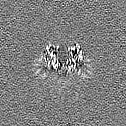
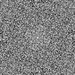
| ファイル | ダウンロード / ファイル: emd_8912.map.gz / 形式: CCP4 / 大きさ: 64 MB / タイプ: IMAGE STORED AS FLOATING POINT NUMBER (4 BYTES) | ||||||||||||||||||||||||||||||||||||||||||||||||||||||||||||
|---|---|---|---|---|---|---|---|---|---|---|---|---|---|---|---|---|---|---|---|---|---|---|---|---|---|---|---|---|---|---|---|---|---|---|---|---|---|---|---|---|---|---|---|---|---|---|---|---|---|---|---|---|---|---|---|---|---|---|---|---|---|
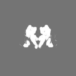
| 注釈 | Human Polycsytin 2-l1, primary map | ||||||||||||||||||||||||||||||||||||||||||||||||||||||||||||
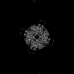
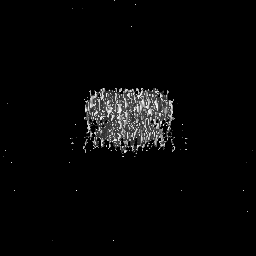
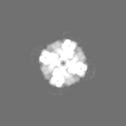
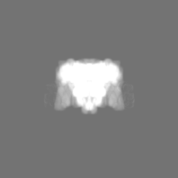
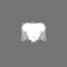
| 投影像・断面図 | 画像のコントロール
画像は Spider により作成 | ||||||||||||||||||||||||||||||||||||||||||||||||||||||||||||
| ボクセルのサイズ | X=Y=Z: 1.04 Å | ||||||||||||||||||||||||||||||||||||||||||||||||||||||||||||
| 密度 |
| ||||||||||||||||||||||||||||||||||||||||||||||||||||||||||||
| 対称性 | 空間群: 1 | ||||||||||||||||||||||||||||||||||||||||||||||||||||||||||||
| 詳細 | EMDB XML:
CCP4マップ ヘッダ情報:
| ||||||||||||||||||||||||||||||||||||||||||||||||||||||||||||
 Z (Sec.)
 Y (Row.)
 X (Col.)


-添付データ
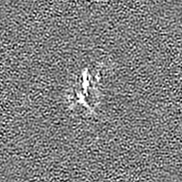
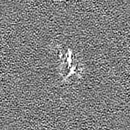
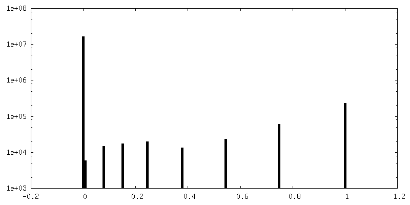
-マスク #1
| ファイル | emd_8912_msk_1.map | ||||||||||||
|---|---|---|---|---|---|---|---|---|---|---|---|---|---|
| 投影像・断面図 |
| ||||||||||||
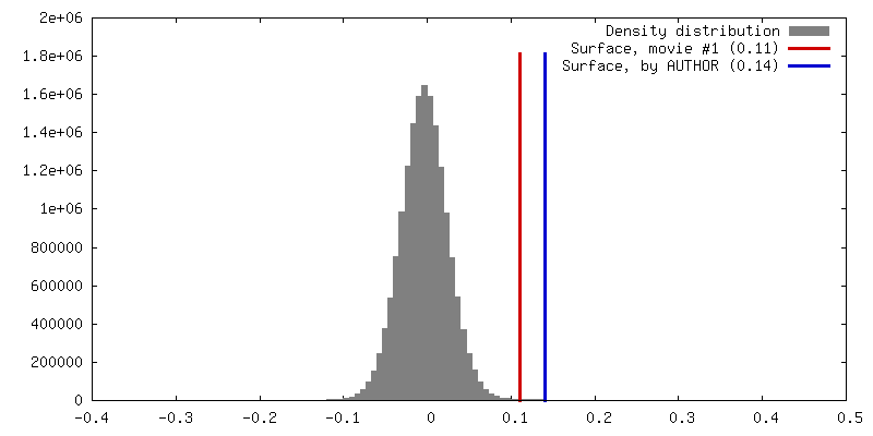
| 密度ヒストグラム |


- 試料の構成要素
-全体 : Human polycystic 2-l
| 全体 | 名称: Human polycystic 2-l |
|---|---|
| 要素 |
|
-超分子 #1: Human polycystic 2-l
| 超分子 | 名称: Human polycystic 2-l / タイプ: organelle_or_cellular_component / ID: 1 / 親要素: 0 / 含まれる分子: #1 / 詳細: Homotetrameric assembly of polycystic 2-l1 |
|---|---|
| 由来(天然) | 生物種: Homo sapiens (ヒト) |
| 分子量 | 理論値: 547 KDa |
-分子 #1: Polycystic kidney disease 2-like 1 protein
| 分子 | 名称: Polycystic kidney disease 2-like 1 protein / タイプ: protein_or_peptide / ID: 1 / コピー数: 4 / 光学異性体: LEVO |
|---|---|
| 由来(天然) | 生物種: Homo sapiens (ヒト) |
| 分子量 | 理論値: 92.070633 KDa |
| 組換発現 | 生物種: Homo sapiens (ヒト) |
| 配列 | 文字列: MNAVGSPEGQ ELQKLGSGAW DNPAYSGPPS PHGTLRVCTI SSTGPLQPQP KKPEDEPQET AYRTQVSSCC LHICQGIRGL WGTTLTENT AENRELYIKT TLRELLVYIV FLVDICLLTY GMTSSSAYYY TKVMSELFLH TPSDTGVSFQ AISSMADFWD F AQGPLLDS ...文字列: MNAVGSPEGQ ELQKLGSGAW DNPAYSGPPS PHGTLRVCTI SSTGPLQPQP KKPEDEPQET AYRTQVSSCC LHICQGIRGL WGTTLTENT AENRELYIKT TLRELLVYIV FLVDICLLTY GMTSSSAYYY TKVMSELFLH TPSDTGVSFQ AISSMADFWD F AQGPLLDS LYWTKWYNNQ SLGHGSHSFI YYENMLLGVP RLRQLKVRND SCVVHEDFRE DILSCYDVYS PDKEEQLPFG PF NGTAWTY HSQDELGGFS HWGRLTSYSG GGYYLDLPGS RQGSAEALRA LQEGLWLDRG TRVVFIDFSV YNANINLFCV LRL VVEFPA TGGAIPSWQI RTVKLIRYVS NWDFFIVGCE VIFCVFIFYY VVEEILELHI HRLRYLSSIW NILDLVVILL SIVA VGFHI FRTLEVNRLM GKLLQQPNTY ADFEFLAFWQ TQYNNMNAVN LFFAWIKIFK YISFNKTMTQ LSSTLARCAK DILGF AVMF FIVFFAYAQL GYLLFGTQVE NFSTFIKCIF TQFRIILGDF DYNAIDNANR ILGPAYFVTY VFFVFFVLLN MFLAII NDT YSEVKEELAG QKDELQLSDL LKQGYNKTLL RLRLRKERVS DVQKVLQGGE QEIQFEDFTN TLRELGHAEH EITELTA TF TKFDRDGNRI LDEKEQEKMR QDLEEERVAL NTEIEKLGRS IVSSPQGKSG PEAARAGGWV SGEEFYMLTR RVLQLETV L EGVVSQIDAV GSKLKMLERK GWLAPSPGVK EQAIWKHPQP APAVTPDPWG VQGGQESEVP YKREEEALEE RRLSRGEIP TLQRS UniProtKB: Polycystin-2-like protein 1 |
-分子 #2: 2-acetamido-2-deoxy-beta-D-glucopyranose
| 分子 | 名称: 2-acetamido-2-deoxy-beta-D-glucopyranose / タイプ: ligand / ID: 2 / コピー数: 4 / 式: NAG |
|---|---|
| 分子量 | 理論値: 221.208 Da |
| Chemical component information |  ChemComp-NAG: |
-実験情報
-構造解析
| 手法 | クライオ電子顕微鏡法 |
|---|---|
 解析 解析 | 単粒子再構成法 |
| 試料の集合状態 | 3D array |
-試料調製
| 濃度 | 3.5 mg/mL | |||||||||||||||
|---|---|---|---|---|---|---|---|---|---|---|---|---|---|---|---|---|
| 緩衝液 | pH: 7.5 構成要素:
| |||||||||||||||
| グリッド | モデル: Quantifoil R1.2/1.3 / 材質: COPPER / メッシュ: 400 / 支持フィルム - 材質: FORMVAR / 支持フィルム - トポロジー: HOLEY / 前処理 - タイプ: GLOW DISCHARGE / 前処理 - 時間: 60 sec. | |||||||||||||||
| 凍結 | 凍結剤: ETHANE / チャンバー内湿度: 100 % / チャンバー内温度: 277.15 K / 装置: FEI VITROBOT MARK IV | |||||||||||||||
| 詳細 | polycystic 2-l1 stabilized in amphipol PMAL-C8 |
- 電子顕微鏡法
| 顕微鏡 | FEI TITAN KRIOS |
|---|---|
| 撮影 | フィルム・検出器のモデル: GATAN K2 SUMMIT (4k x 4k) 平均電子線量: 60.0 e/Å2 |
| 電子線 | 加速電圧: 300 kV / 電子線源:  FIELD EMISSION GUN FIELD EMISSION GUN |
| 電子光学系 | 照射モード: OTHER / 撮影モード: OTHER |
| 実験機器 |  モデル: Titan Krios / 画像提供: FEI Company |