+ Open data
- Basic information
| Entry | Database: EMDB / ID: EMD-8837 | |||||||||
|---|---|---|---|---|---|---|---|---|---|---|
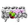
| Title | Sub-tomogram average of I1 dynein Post class | |||||||||
 Map data Map data | Sub-tomogram average of I1 dynein Post class: the two dynein heavy chains of sea urchin sperm flagellar I1 dynein complex are in the post-powerstroke state. | |||||||||
 Sample Sample |
| |||||||||
| Biological species |  | |||||||||
| Method | subtomogram averaging / cryo EM / Resolution: 41.0 Å | |||||||||
 Authors Authors | Lin J / Nicastro D | |||||||||
| Funding support |  United States, 2 items United States, 2 items
| |||||||||
 Citation Citation |  Journal: Science / Year: 2018 Journal: Science / Year: 2018Title: Asymmetric distribution and spatial switching of dynein activity generates ciliary motility. Authors: Jianfeng Lin / Daniela Nicastro /  Abstract: Motile cilia and flagella are essential, highly conserved organelles, and their motility is driven by the coordinated activities of multiple dynein isoforms. The prevailing "switch-point" hypothesis ...Motile cilia and flagella are essential, highly conserved organelles, and their motility is driven by the coordinated activities of multiple dynein isoforms. The prevailing "switch-point" hypothesis posits that dyneins are asymmetrically activated to drive flagellar bending. To test this model, we applied cryo-electron tomography to visualize activity states of individual dyneins relative to their locations along beating flagella of sea urchin sperm cells. As predicted, bending was generated by the asymmetric distribution of dynein activity on opposite sides of the flagellum. However, contrary to predictions, most dyneins were in their active state, and the smaller population of conformationally inactive dyneins switched flagellar sides relative to the bending direction. Thus, our data suggest a "switch-inhibition" mechanism in which force imbalance is generated by inhibiting, rather than activating, dyneins on alternating sides of the flagellum. | |||||||||
| History |
|
- Structure visualization
| Movie |
 Movie viewer Movie viewer |
|---|---|
| Structure viewer | EM map:  SurfView SurfView Molmil Molmil Jmol/JSmol Jmol/JSmol |
| Supplemental images |
- Downloads & links
-EMDB archive
| Map data |  emd_8837.map.gz emd_8837.map.gz | 591.9 KB |  EMDB map data format EMDB map data format | |
|---|---|---|---|---|
| Header (meta data) |  emd-8837-v30.xml emd-8837-v30.xml emd-8837.xml emd-8837.xml | 11.2 KB 11.2 KB | Display Display |  EMDB header EMDB header |
| Images |  emd_8837.png emd_8837.png | 85.5 KB | ||
| Archive directory |  http://ftp.pdbj.org/pub/emdb/structures/EMD-8837 http://ftp.pdbj.org/pub/emdb/structures/EMD-8837 ftp://ftp.pdbj.org/pub/emdb/structures/EMD-8837 ftp://ftp.pdbj.org/pub/emdb/structures/EMD-8837 | HTTPS FTP |
-Validation report
| Summary document |  emd_8837_validation.pdf.gz emd_8837_validation.pdf.gz | 78.1 KB | Display |  EMDB validaton report EMDB validaton report |
|---|---|---|---|---|
| Full document |  emd_8837_full_validation.pdf.gz emd_8837_full_validation.pdf.gz | 77.2 KB | Display | |
| Data in XML |  emd_8837_validation.xml.gz emd_8837_validation.xml.gz | 494 B | Display | |
| Arichive directory |  https://ftp.pdbj.org/pub/emdb/validation_reports/EMD-8837 https://ftp.pdbj.org/pub/emdb/validation_reports/EMD-8837 ftp://ftp.pdbj.org/pub/emdb/validation_reports/EMD-8837 ftp://ftp.pdbj.org/pub/emdb/validation_reports/EMD-8837 | HTTPS FTP |
-Related structure data
| Related structure data |  8835C  8836C  8838C C: citing same article ( |
|---|---|
| Similar structure data | |
| EM raw data |  EMPIAR-10158 (Title: Cryo electron tomography of immotile sea urchin sperm flagella EMPIAR-10158 (Title: Cryo electron tomography of immotile sea urchin sperm flagellaData size: 13.8 Data #1: Tilt series of immotile sea urchin sperm flagella [tilt series]) |
- Links
Links
| EMDB pages |  EMDB (EBI/PDBe) / EMDB (EBI/PDBe) /  EMDataResource EMDataResource |
|---|
- Map
Map
| File |  Download / File: emd_8837.map.gz / Format: CCP4 / Size: 704.1 KB / Type: IMAGE STORED AS FLOATING POINT NUMBER (4 BYTES) Download / File: emd_8837.map.gz / Format: CCP4 / Size: 704.1 KB / Type: IMAGE STORED AS FLOATING POINT NUMBER (4 BYTES) | ||||||||||||||||||||||||||||||||||||||||||||||||||||||||||||||||||||
|---|---|---|---|---|---|---|---|---|---|---|---|---|---|---|---|---|---|---|---|---|---|---|---|---|---|---|---|---|---|---|---|---|---|---|---|---|---|---|---|---|---|---|---|---|---|---|---|---|---|---|---|---|---|---|---|---|---|---|---|---|---|---|---|---|---|---|---|---|---|
| Annotation | Sub-tomogram average of I1 dynein Post class: the two dynein heavy chains of sea urchin sperm flagellar I1 dynein complex are in the post-powerstroke state. | ||||||||||||||||||||||||||||||||||||||||||||||||||||||||||||||||||||
| Projections & slices | Image control
Images are generated by Spider. generated in cubic-lattice coordinate | ||||||||||||||||||||||||||||||||||||||||||||||||||||||||||||||||||||
| Voxel size | X=Y=Z: 9.856 Å | ||||||||||||||||||||||||||||||||||||||||||||||||||||||||||||||||||||
| Density |
| ||||||||||||||||||||||||||||||||||||||||||||||||||||||||||||||||||||
| Symmetry | Space group: 1 | ||||||||||||||||||||||||||||||||||||||||||||||||||||||||||||||||||||
| Details | EMDB XML:
CCP4 map header:
| ||||||||||||||||||||||||||||||||||||||||||||||||||||||||||||||||||||
-Supplemental data
- Sample components
-Entire : Sea urchin sperm
| Entire | Name: Sea urchin sperm |
|---|---|
| Components |
|
-Supramolecule #1: Sea urchin sperm
| Supramolecule | Name: Sea urchin sperm / type: cell / ID: 1 / Parent: 0 / Macromolecule list: #1 Details: Sperm were diluted in demembranation buffer (30 mM HEPES, pH 8.0, 150 mM KCl, 4 mM MgCl2, 0.5 mM EGTA, 0.1% Triton X-100) and incubated for one minute to remove the flagellar membrane. The ...Details: Sperm were diluted in demembranation buffer (30 mM HEPES, pH 8.0, 150 mM KCl, 4 mM MgCl2, 0.5 mM EGTA, 0.1% Triton X-100) and incubated for one minute to remove the flagellar membrane. The sperm were then collected by centrifugation at 1000x g, and resuspended in demembranation buffer (but without Triton X-100). |
|---|---|
| Source (natural) | Organism:  |
-Experimental details
-Structure determination
| Method | cryo EM |
|---|---|
 Processing Processing | subtomogram averaging |
| Aggregation state | cell |
- Sample preparation
| Buffer | pH: 8 Component:
| |||||||||||||||
|---|---|---|---|---|---|---|---|---|---|---|---|---|---|---|---|---|
| Grid | Model: Quantifoil R2/2 / Material: COPPER / Mesh: 200 / Support film - Material: CARBON / Support film - topology: HOLEY / Pretreatment - Type: GLOW DISCHARGE | |||||||||||||||
| Vitrification | Cryogen name: ETHANE / Instrument: HOMEMADE PLUNGER |
- Electron microscopy
| Microscope | FEI TECNAI F30 |
|---|---|
| Specialist optics | Energy filter - Name: GIF |
| Image recording | Film or detector model: GATAN MULTISCAN / Average electron dose: 1.5 e/Å2 |
| Electron beam | Acceleration voltage: 300 kV / Electron source:  FIELD EMISSION GUN FIELD EMISSION GUN |
| Electron optics | C2 aperture diameter: 100.0 µm / Illumination mode: FLOOD BEAM / Imaging mode: BRIGHT FIELD / Nominal defocus max: 8.0 µm / Nominal defocus min: 6.0 µm / Nominal magnification: 13500 |
| Sample stage | Cooling holder cryogen: NITROGEN |
| Experimental equipment |  Model: Tecnai F30 / Image courtesy: FEI Company |
- Image processing
| Final reconstruction | Resolution.type: BY AUTHOR / Resolution: 41.0 Å / Resolution method: FSC 0.5 CUT-OFF / Software - Name: PEET (ver. 1.9.0) / Number subtomograms used: 392 |
|---|---|
| Extraction | Number tomograms: 7 / Number images used: 1134 / Software - Name: PEET (ver. 1.9.0) |
| Final 3D classification | Software - Name: PEET (ver. 1.9.0) |
| Final angle assignment | Type: OTHER |
 Movie
Movie Controller


 Z (Sec.)
 Y (Row.)
 X (Col.)


