regulation of synaptic vesicle docking / glutamatergic postsynaptic density / HSF1-dependent transactivation / RAF activation / Ion transport by P-type ATPases / peptidyl-threonine autophosphorylation / regulation of endocannabinoid signaling pathway / neurotransmitter receptor transport to plasma membrane / : / calcium- and calmodulin-dependent protein kinase complex ...regulation of synaptic vesicle docking / glutamatergic postsynaptic density / HSF1-dependent transactivation / RAF activation / Ion transport by P-type ATPases / peptidyl-threonine autophosphorylation / regulation of endocannabinoid signaling pathway / neurotransmitter receptor transport to plasma membrane / : / calcium- and calmodulin-dependent protein kinase complex / Interferon gamma signaling / regulation of neuron migration / Ca2+/calmodulin-dependent protein kinase / regulation of neurotransmitter secretion / calcium-dependent protein serine/threonine kinase activity / dendritic spine development / Trafficking of AMPA receptors / Ca2+ pathway / positive regulation of calcium ion transport / presynaptic cytosol / negative regulation of hydrolase activity / postsynaptic neurotransmitter receptor diffusion trapping / postsynaptic specialization membrane / GTPase activating protein binding / RAF/MAP kinase cascade / dendrite morphogenesis / NMDA selective glutamate receptor signaling pathway / regulation of mitochondrial membrane permeability involved in apoptotic process / regulation of neurotransmitter receptor localization to postsynaptic specialization membrane / postsynaptic cytosol / calmodulin-dependent protein kinase activity / Ion homeostasis / positive regulation of cardiac muscle cell apoptotic process / regulation of neuronal synaptic plasticity / Unblocking of NMDA receptors, glutamate binding and activation / cellular response to interferon-beta / regulation of protein localization to plasma membrane / glutamate receptor binding / ionotropic glutamate receptor signaling pathway / dendrite cytoplasm / response to ischemia / angiotensin-activated signaling pathway / positive regulation of receptor signaling pathway via JAK-STAT / Schaffer collateral - CA1 synapse / cellular response to type II interferon / G1/S transition of mitotic cell cycle / calcium ion transport / kinase activity / dendritic spine / postsynaptic density / calmodulin binding / neuron projection / axon / protein phosphorylation / protein serine kinase activity / protein serine/threonine kinase activity / neuronal cell body / glutamatergic synapse / synapse / dendrite / protein homodimerization activity / mitochondrion / ATP binding / identical protein binding / metal ion binding / cytoplasm / cytosol Similarity search - Function
Calcium/calmodulin-dependent protein kinase II, association-domain / Calcium/calmodulin dependent protein kinase II association domain / NTF2-like domain superfamily / Serine/threonine-protein kinase, active site / Serine/Threonine protein kinases active-site signature. / Protein kinase domain / Serine/Threonine protein kinases, catalytic domain / Protein kinase, ATP binding site / Protein kinases ATP-binding region signature. / Protein kinase domain profile. ...Calcium/calmodulin-dependent protein kinase II, association-domain / Calcium/calmodulin dependent protein kinase II association domain / NTF2-like domain superfamily / Serine/threonine-protein kinase, active site / Serine/Threonine protein kinases active-site signature. / Protein kinase domain / Serine/Threonine protein kinases, catalytic domain / Protein kinase, ATP binding site / Protein kinases ATP-binding region signature. / Protein kinase domain profile. / Protein kinase domain / Protein kinase-like domain superfamily Similarity search - Domain/homology
National Institutes of Health/National Institute of Neurological Disorders and Stroke (NIH/NINDS)
R01NS081248
United States
Citation
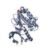
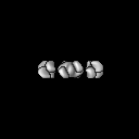
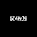
Journal: Nat Commun / Year: 2017 Title: The CaMKII holoenzyme structure in activation-competent conformations. Authors: Janette B Myers / Vincent Zaegel / Steven J Coultrap / Adam P Miller / K Ulrich Bayer / Steve L Reichow / Abstract: The Ca/calmodulin-dependent protein kinase II (CaMKII) assembles into large 12-meric holoenzymes, which is thought to enable regulatory processes required for synaptic plasticity underlying learning, ...The Ca/calmodulin-dependent protein kinase II (CaMKII) assembles into large 12-meric holoenzymes, which is thought to enable regulatory processes required for synaptic plasticity underlying learning, memory and cognition. Here we used single particle electron microscopy (EM) to determine a pseudoatomic model of the CaMKIIα holoenzyme in an extended and activation-competent conformation. The holoenzyme is organized by a rigid central hub complex, while positioning of the kinase domains is highly flexible, revealing dynamic holoenzymes ranging from 15-35 nm in diameter. While most kinase domains are ordered independently, ∼20% appear to form dimers and <3% are consistent with a compact conformation. An additional level of plasticity is revealed by a small fraction of bona-fide 14-mers (<4%) that may enable subunit exchange. Biochemical and cellular FRET studies confirm that the extended state of CaMKIIα resolved by EM is the predominant form of the holoenzyme, even under molecular crowding conditions.
History
Deposition
Dec 9, 2016
-
Header (metadata) release
Dec 28, 2016
-
Map release
Jun 21, 2017
-
Update
Mar 13, 2024
-
Current status
Mar 13, 2024
Processing site: RCSB / Status: Released
-
Structure visualization
Movie
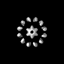
Surface view with section colored by density value
Entire : Calcium-calmodulin dependent kinase II alpha
Entire
Name: Calcium-calmodulin dependent kinase II alpha
Components
Complex: Calcium-calmodulin dependent kinase II alpha
Protein or peptide: Calcium/calmodulin-dependent protein kinase type II subunit alpha
-
Supramolecule #1: Calcium-calmodulin dependent kinase II alpha
Supramolecule
Name: Calcium-calmodulin dependent kinase II alpha / type: complex / ID: 1 / Parent: 0 / Macromolecule list: all / Details: Full-length CaMKII alpha wild type
Source (natural)
Organism: Rattus norvegicus (Norway rat)
Molecular weight
Theoretical: 620 KDa
-
Macromolecule #1: Calcium/calmodulin-dependent protein kinase type II subunit alpha
Macromolecule
Name: Calcium/calmodulin-dependent protein kinase type II subunit alpha type: protein_or_peptide / ID: 1 / Number of copies: 12 / Enantiomer: LEVO / EC number: Ca2+/calmodulin-dependent protein kinase
Number classes used: 1 / Applied symmetry - Point group: D6 (2x6 fold dihedral) / Resolution.type: BY AUTHOR / Resolution: 20.0 Å / Resolution method: FSC 0.143 CUT-OFF / Software - Name: RELION (ver. 1.4) Details: Only modeled the unstructured linker region, residues 300-345. The rest came from two other, previously published structures, namely 5IG3 and 2VZ6 Number images used: 2000
Initial angle assignment
Type: RANDOM ASSIGNMENT / Software - Name: EMAN (ver. 2.12)
Final angle assignment
Type: OTHER / Software - Name: RELION (ver. 1.4)
Final 3D classification
Number classes: 6 / Software - Name: RELION (ver. 1.4)
In the structure databanks used in Yorodumi, some data are registered as the other names, "COVID-19 virus" and "2019-nCoV". Here are the details of the virus and the list of structure data.
Jan 31, 2019. EMDB accession codes are about to change! (news from PDBe EMDB page)
EMDB accession codes are about to change! (news from PDBe EMDB page)
The allocation of 4 digits for EMDB accession codes will soon come to an end. Whilst these codes will remain in use, new EMDB accession codes will include an additional digit and will expand incrementally as the available range of codes is exhausted. The current 4-digit format prefixed with “EMD-” (i.e. EMD-XXXX) will advance to a 5-digit format (i.e. EMD-XXXXX), and so on. It is currently estimated that the 4-digit codes will be depleted around Spring 2019, at which point the 5-digit format will come into force.
The EM Navigator/Yorodumi systems omit the EMD- prefix.
Related info.:Q: What is EMD? / ID/Accession-code notation in Yorodumi/EM Navigator
Yorodumi is a browser for structure data from EMDB, PDB, SASBDB, etc.
This page is also the successor to EM Navigator detail page, and also detail information page/front-end page for Omokage search.
The word "yorodu" (or yorozu) is an old Japanese word meaning "ten thousand". "mi" (miru) is to see.
Related info.:EMDB / PDB / SASBDB / Comparison of 3 databanks / Yorodumi Search / Aug 31, 2016. New EM Navigator & Yorodumi / Yorodumi Papers / Jmol/JSmol / Function and homology information / Changes in new EM Navigator and Yorodumi
 Movie
Movie Controller


 Open data
 Basic information
 Map data
 Sample
 Keywords
 Function and homology information
 Authors
 United States, 1 items
  Citation
 Structure visualization
 UCSF Chimera
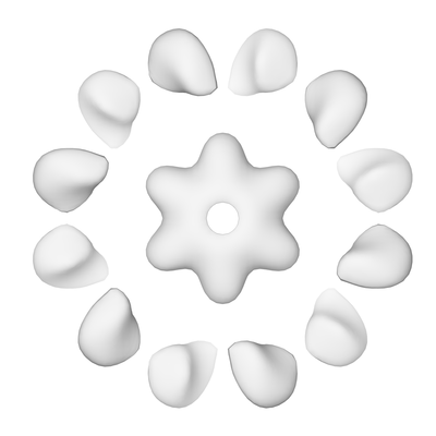
 Downloads & links
 emd_8514.png
 http://ftp.pdbj.org/pub/emdb/structures/EMD-8514


 Z (Sec.)
 Y (Row.)
 X (Col.)


 Sample components
 Spodoptera frugiperda (fall armyworm)
 Processing
 Electron microscopy