Movie
Movie Controller


+ Open data

- Basic information
| Entry | Database: PDB / ID: 5u6y | ||||||
|---|---|---|---|---|---|---|---|
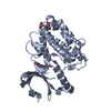
| Title | Pseudo-atomic model of the CaMKIIa holoenzyme. | ||||||
 Components Components | Calcium/calmodulin-dependent protein kinase type II subunit alpha | ||||||
 Keywords Keywords | TRANSFERASE / Calcium/calmodulin-dependent kinase II (CaMKII) / cell signaling / calcium / calmodulin (CaM) / long-term potentiation (LTP) / long-term depression (LTD) / synaptic plasticity / cooperativity / electron microscopy (EM) / single particle reconstruction / intrinsic disorder | ||||||
| Function / homology |  Function and homology information Function and homology information: / HSF1-dependent transactivation / RAF activation / Ion transport by P-type ATPases / peptidyl-threonine autophosphorylation / calcium- and calmodulin-dependent protein kinase complex / regulation of endocannabinoid signaling pathway / neurotransmitter receptor transport to plasma membrane / Interferon gamma signaling / Ca2+/calmodulin-dependent protein kinase ...: / HSF1-dependent transactivation / RAF activation / Ion transport by P-type ATPases / peptidyl-threonine autophosphorylation / calcium- and calmodulin-dependent protein kinase complex / regulation of endocannabinoid signaling pathway / neurotransmitter receptor transport to plasma membrane / Interferon gamma signaling / Ca2+/calmodulin-dependent protein kinase / dendritic spine development / negative regulation of hydrolase activity / regulation of neurotransmitter secretion / Trafficking of AMPA receptors / positive regulation of calcium ion transport / regulation of neuron migration / calcium/calmodulin-dependent protein kinase activity / Ca2+ pathway / regulation of mitochondrial membrane permeability involved in apoptotic process / RAF/MAP kinase cascade / GTPase activating protein binding / Ion homeostasis / dendrite morphogenesis / regulation of neurotransmitter receptor localization to postsynaptic specialization membrane / negative regulation of ferroptosis / Unblocking of NMDA receptors, glutamate binding and activation / regulation of neuronal synaptic plasticity / glutamate receptor binding / postsynaptic cytosol / cell surface receptor signaling pathway via JAK-STAT / regulation of protein localization to plasma membrane / presynaptic cytosol / cellular response to interferon-beta / positive regulation of cardiac muscle cell apoptotic process / ionotropic glutamate receptor signaling pathway / dendrite cytoplasm / response to ischemia / angiotensin-activated signaling pathway / positive regulation of receptor signaling pathway via JAK-STAT / G1/S transition of mitotic cell cycle / cellular response to type II interferon / Schaffer collateral - CA1 synapse / kinase activity / calcium ion transport / dendritic spine / calmodulin binding / neuron projection / postsynaptic density / protein serine kinase activity / axon / protein serine/threonine kinase activity / neuronal cell body / synapse / dendrite / glutamatergic synapse / protein homodimerization activity / mitochondrion / ATP binding / metal ion binding / identical protein binding / cytoplasm / cytosol Similarity search - Function | ||||||
| Biological species |  | ||||||
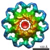
| Method | ELECTRON MICROSCOPY / single particle reconstruction / negative staining / Resolution: 20 Å | ||||||
 Authors Authors | Myers, J. / Reichow, S.L. | ||||||
| Funding support |  United States, 1items United States, 1items
| ||||||
 Citation Citation | Journal: Nat Commun / Year: 2017 Title: The CaMKII holoenzyme structure in activation-competent conformations. Authors: Janette B Myers / Vincent Zaegel / Steven J Coultrap / Adam P Miller / K Ulrich Bayer / Steve L Reichow / Abstract: The Ca/calmodulin-dependent protein kinase II (CaMKII) assembles into large 12-meric holoenzymes, which is thought to enable regulatory processes required for synaptic plasticity underlying learning, ...The Ca/calmodulin-dependent protein kinase II (CaMKII) assembles into large 12-meric holoenzymes, which is thought to enable regulatory processes required for synaptic plasticity underlying learning, memory and cognition. Here we used single particle electron microscopy (EM) to determine a pseudoatomic model of the CaMKIIα holoenzyme in an extended and activation-competent conformation. The holoenzyme is organized by a rigid central hub complex, while positioning of the kinase domains is highly flexible, revealing dynamic holoenzymes ranging from 15-35 nm in diameter. While most kinase domains are ordered independently, ∼20% appear to form dimers and <3% are consistent with a compact conformation. An additional level of plasticity is revealed by a small fraction of bona-fide 14-mers (<4%) that may enable subunit exchange. Biochemical and cellular FRET studies confirm that the extended state of CaMKIIα resolved by EM is the predominant form of the holoenzyme, even under molecular crowding conditions. | ||||||
| History |
|
- Structure visualization
| Movie |
Movie viewer |
|---|---|
| Structure viewer | Molecule: MolmilJmol/JSmol |
 UCSF Chimera
- Downloads & links
-Download
| PDBx/mmCIF format | 5u6y.cif.gz | 1.8 MB | Display | PDBx/mmCIF format |
|---|---|---|---|---|
| PDB format | pdb5u6y.ent.gz | 1.6 MB | Display | PDB format |
| PDBx/mmJSON format | 5u6y.json.gz | Tree view | PDBx/mmJSON format | |
| Others |  Other downloads Other downloads |
-Validation report
| Arichive directory | https://data.pdbj.org/pub/pdb/validation_reports/u6/5u6yftp://data.pdbj.org/pub/pdb/validation_reports/u6/5u6y | HTTPS FTP |
|---|
-Related structure data
| Related structure data | 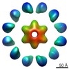 8514MC M: map data used to model this data C: citing same article ( |
|---|---|
| Similar structure data |
-Links
 PDBj
PDBj


- Assembly
| Deposited unit | 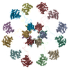
|
|---|---|
| 1 |
|
-Components
| #1: Protein | Mass: 52281.535 Da / Num. of mol.: 12 Source method: isolated from a genetically manipulated source Source: (gene. exp.)   Spodoptera frugiperda (fall armyworm) Spodoptera frugiperda (fall armyworm)References: UniProt: P11275, Ca2+/calmodulin-dependent protein kinase |
|---|
-Experimental details
-Experiment
| Experiment | Method: ELECTRON MICROSCOPY |
|---|---|
| EM experiment | Aggregation state: PARTICLE / 3D reconstruction method: single particle reconstruction |
- Sample preparation
| Component | Name: Calcium-calmodulin dependent kinase II alpha / Type: COMPLEX / Details: Full-length CaMKII alpha wild type / Entity ID: all / Source: RECOMBINANT | ||||||||||||||||||||
|---|---|---|---|---|---|---|---|---|---|---|---|---|---|---|---|---|---|---|---|---|---|
| Molecular weight | Value: 0.62 MDa / Experimental value: NO | ||||||||||||||||||||
| Source (natural) | Organism: | ||||||||||||||||||||
| Source (recombinant) | Organism: Spodoptera frugiperda (fall armyworm) / Plasmid: pFastBac1 | ||||||||||||||||||||
| Buffer solution | pH: 7.4 | ||||||||||||||||||||
| Buffer component |
| ||||||||||||||||||||
| Specimen | Embedding applied: NO / Shadowing applied: NO / Staining applied: YES / Vitrification applied: NO / Details: 100 nM complex | ||||||||||||||||||||
| EM staining | Type: NEGATIVE / Details: 0.75% (wt/vol) uranyl formate / Material: Uranyl Formate | ||||||||||||||||||||
| Specimen support | Grid material: COPPER / Grid mesh size: 200 divisions/in. / Grid type: Ted Pella |
- Electron microscopy imaging
| Microscopy | Model: FEI TECNAI 12 |
|---|---|
| Electron gun | Electron source: TUNGSTEN HAIRPIN / Accelerating voltage: 120 kV / Illumination mode: FLOOD BEAM |
| Electron lens | Mode: BRIGHT FIELD / Nominal magnification: 49000 X |
| Image recording | Average exposure time: 1 sec. / Electron dose: 20 e/Å2 / Film or detector model: FEI EAGLE (2k x 2k) |
- Processing
| EM software |
| ||||||||||||||||||||||||||||||||||||||||
|---|---|---|---|---|---|---|---|---|---|---|---|---|---|---|---|---|---|---|---|---|---|---|---|---|---|---|---|---|---|---|---|---|---|---|---|---|---|---|---|---|---|
| CTF correction | Details: Performed in EMAN2 / Type: PHASE FLIPPING AND AMPLITUDE CORRECTION | ||||||||||||||||||||||||||||||||||||||||
| Particle selection | Num. of particles selected: 16616 | ||||||||||||||||||||||||||||||||||||||||
| Symmetry | Point symmetry: D6 (2x6 fold dihedral) | ||||||||||||||||||||||||||||||||||||||||
| 3D reconstruction | Resolution: 20 Å / Resolution method: FSC 0.143 CUT-OFF / Num. of particles: 2000 Details: Only modeled the unstructured linker region, residues 300-345. The rest came from two other, previously published structures, namely 5IG3 and 2VZ6 Num. of class averages: 1 / Symmetry type: POINT | ||||||||||||||||||||||||||||||||||||||||
| Atomic model building | Protocol: RIGID BODY FIT / Target criteria: Correlation Coefficient | ||||||||||||||||||||||||||||||||||||||||
| Atomic model building | 3D fitting-ID: 1 / Source name: PDB / Type: experimental model
|