 Movie
Movie Controller


+ Open data

- Basic information
| Entry |  | |||||||||
|---|---|---|---|---|---|---|---|---|---|---|
| Title | Cryo-EM structure of cUA bound CapE filament | |||||||||
 Map data Map data | ||||||||||
 Sample Sample |
| |||||||||
 Keywords Keywords | CapE / patatin-like phospholipase protein / LIPID BINDING PROTEIN | |||||||||
| Function / homology |  Function and homology information Function and homology informationphospholipase A1 / phospholipase A1 activity / lipid catabolic process / fatty acid metabolic process / defense response to virus / membrane Similarity search - Function | |||||||||
| Biological species |  | |||||||||
| Method | single particle reconstruction / cryo EM / Resolution: 3.27 Å | |||||||||
 Authors Authors | Gao A / Wang JG | |||||||||
| Funding support | 1 items
| |||||||||
 Citation Citation | Journal: Cell / Year: 2025 Title: Cyclic-dinucleotide-induced filamentous assembly of phospholipases governs broad CBASS immunity. Authors: Jingge Wang / Zhao Li / Hao Lang / Wenfeng Fu / Yina Gao / Sen Yin / Panpan Sun / Zhaolong Li / Jiafeng Huang / Songqing Liu / Yun Zhu / Fei Sun / Dong Li / Pu Gao / Ang Gao /  Abstract: Cyclic-oligonucleotide-based antiphage signaling systems (CBASS), a widespread antiviral bacterial immune system homologous to the mammalian cGAS-STING pathway, synthesizes cyclic nucleotide signals ...Cyclic-oligonucleotide-based antiphage signaling systems (CBASS), a widespread antiviral bacterial immune system homologous to the mammalian cGAS-STING pathway, synthesizes cyclic nucleotide signals and triggers effector proteins to induce cell death and prevent viral propagation. Among various CBASS effectors, phospholipase effectors are the first to be discovered and are one of the most widespread families that sense cyclic dinucleotides to degrade cell membrane phospholipids. Here, we report that CBASS phospholipases assemble from a dimeric inactive state into active higher-order filamentous oligomers upon sensing cyclic dinucleotides. Using a combined approach of cryo-electron microscopy and X-ray crystallography, we have determined the structures of CBASS phospholipase in the inactive dimeric state, the cyclic-dinucleotide-bound active higher-order state, and the substrate-analog-bound catalytic mimicry state, thereby visualizing the complete conformational reorganization process. Complemented by functional assays of intermolecular binding, phospholipase enzymatic activity, in vitro membrane disruption, and in vivo antiphage efficiency, our work elucidates the mechanisms of assembly and activation of CBASS phospholipases. | |||||||||
| History |
|
- Structure visualization
| Supplemental images |
|---|

- Downloads & links
-EMDB archive
| Map data | emd_60745.map.gz | 54.5 MB | EMDB map data format | |
|---|---|---|---|---|
| Header (meta data) | emd-60745-v30.xmlemd-60745.xml | 14 KB 14 KB | Display Display | EMDB header |
| FSC (resolution estimation) | emd_60745_fsc.xml | 8.8 KB | Display | FSC data file |
| Images |  emd_60745.png emd_60745.png | 16.8 KB | ||
| Filedesc metadata | emd-60745.cif.gz | 5.6 KB | ||
| Others | emd_60745_half_map_1.map.gzemd_60745_half_map_2.map.gz | 45.2 MB 45.2 MB | ||
| Archive directory |  http://ftp.pdbj.org/pub/emdb/structures/EMD-60745ftp://ftp.pdbj.org/pub/emdb/structures/EMD-60745 http://ftp.pdbj.org/pub/emdb/structures/EMD-60745ftp://ftp.pdbj.org/pub/emdb/structures/EMD-60745 | HTTPS FTP |
-Validation report
| Summary document | emd_60745_validation.pdf.gz | 910.5 KB | Display | EMDB validaton report |
|---|---|---|---|---|
| Full document | emd_60745_full_validation.pdf.gz | 910.1 KB | Display | |
| Data in XML | emd_60745_validation.xml.gz | 14.4 KB | Display | |
| Data in CIF | emd_60745_validation.cif.gz | 20.5 KB | Display | |
| Arichive directory | https://ftp.pdbj.org/pub/emdb/validation_reports/EMD-60745ftp://ftp.pdbj.org/pub/emdb/validation_reports/EMD-60745 | HTTPS FTP |
-Related structure data
| Related structure data |  9ionMC  9iomC 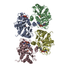 9iopC  9ioqC M: atomic model generated by this map C: citing same article ( |
|---|---|
| Similar structure data |

-Links
| EMDB pages | EMDB (EBI/PDBe) / EMDataResource |
|---|---|
| Related items in Molecule of the Month |

-Map
| File | Download / File: emd_60745.map.gz / Format: CCP4 / Size: 58.2 MB / Type: IMAGE STORED AS FLOATING POINT NUMBER (4 BYTES) | ||||||||||||||||||||||||||||||||||||
|---|---|---|---|---|---|---|---|---|---|---|---|---|---|---|---|---|---|---|---|---|---|---|---|---|---|---|---|---|---|---|---|---|---|---|---|---|---|
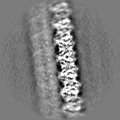
| Projections & slices | Image control
Images are generated by Spider. | ||||||||||||||||||||||||||||||||||||
| Voxel size | X=Y=Z: 1.36 Å | ||||||||||||||||||||||||||||||||||||
| Density |
| ||||||||||||||||||||||||||||||||||||
| Symmetry | Space group: 1 | ||||||||||||||||||||||||||||||||||||
| Details | EMDB XML:
|
 Z (Sec.)
 Y (Row.)
 X (Col.)


-Supplemental data
-Half map: #2
| File | emd_60745_half_map_1.map | ||||||||||||
|---|---|---|---|---|---|---|---|---|---|---|---|---|---|
| Projections & Slices |
| ||||||||||||
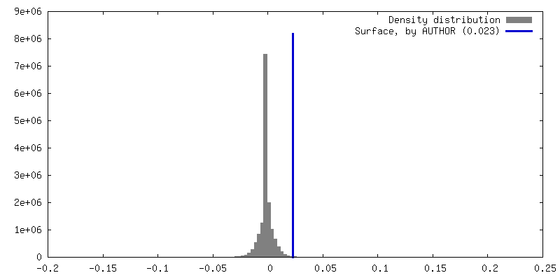
| Density Histograms |


-Half map: #1
| File | emd_60745_half_map_2.map | ||||||||||||
|---|---|---|---|---|---|---|---|---|---|---|---|---|---|
| Projections & Slices |
| ||||||||||||
| Density Histograms |


- Sample components
-Entire : CapE
| Entire | Name: CapE |
|---|---|
| Components |
|
-Supramolecule #1: CapE
| Supramolecule | Name: CapE / type: complex / ID: 1 / Parent: 0 / Macromolecule list: #1 |
|---|---|
| Source (natural) | Organism: |
-Macromolecule #1: cUMP-AMP-activated phospholipase
| Macromolecule | Name: cUMP-AMP-activated phospholipase / type: protein_or_peptide / ID: 1 / Number of copies: 4 / Enantiomer: LEVO / EC number: phospholipase A1 |
|---|---|
| Source (natural) | Organism: |
| Molecular weight | Theoretical: 35.406938 KDa |
| Recombinant expression | Organism: |
| Sequence | String: MTYSVSPSSL LTEYGNDNIC RVLALDGGGA KGFYTLGVLK EIEAMLGCPL YKRFDLVFGT STGAIIAALI ALGYEVDQIH ALYTEHVPR VMSSRSAAAR TMALQDLAKE VFQDKTFEDV LMGIGIVATR WMTERPMIFK GNVVQAHGRK GTFSPGFGVS I ADAVQASC ...String: MTYSVSPSSL LTEYGNDNIC RVLALDGGGA KGFYTLGVLK EIEAMLGCPL YKRFDLVFGT STGAIIAALI ALGYEVDQIH ALYTEHVPR VMSSRSAAAR TMALQDLAKE VFQDKTFEDV LMGIGIVATR WMTERPMIFK GNVVQAHGRK GTFSPGFGVS I ADAVQASC SAYPFFERKV IVTAAGDKVE LIDGGYCANN PTLFAIADAT VALKKDHKDI RVINVGVGIY PEPKPGLLMR IA KKWLAVQ LLQKTLEINT QSMDQLRDIL FKDIPTIRIS DTFERPEMAT DLLEYNLDKL NTLRQRGRES FGAREAQLRE FLI UniProtKB: cUMP-AMP-activated phospholipase |
-Macromolecule #2: 3'3'-cUMP-AMP
| Macromolecule | Name: 3'3'-cUMP-AMP / type: ligand / ID: 2 / Number of copies: 4 / Formula: A1AEP |
|---|---|
| Molecular weight | Theoretical: 635.372 Da |
-Experimental details
-Structure determination
| Method | cryo EM |
|---|---|
 Processing Processing | single particle reconstruction |
| Aggregation state | particle |
-Sample preparation
| Buffer | pH: 8 |
|---|---|
| Vitrification | Cryogen name: ETHANE |
- Electron microscopy
| Microscope | TFS KRIOS |
|---|---|
| Image recording | Film or detector model: GATAN K2 SUMMIT (4k x 4k) / Average electron dose: 60.0 e/Å2 |
| Electron beam | Acceleration voltage: 300 kV / Electron source:  FIELD EMISSION GUN FIELD EMISSION GUN |
| Electron optics | Illumination mode: FLOOD BEAM / Imaging mode: BRIGHT FIELD / Nominal defocus max: 1.5 µm / Nominal defocus min: 1.3 µm |
| Experimental equipment |  Model: Titan Krios / Image courtesy: FEI Company |

