[English] 日本語
Yorodumi
- PDB-9iom: CapE apo form -

+
Open data


ID or keywords:

Loading...

-
Basic information

Entry
Database: PDB / ID: 9iom
TitleCapE apo form
ComponentscUMP-AMP-activated phospholipase
KeywordsLIPID BINDING PROTEIN / CapE / patatin-like phospholipase protein
Function / homology
Function and homology information


phospholipase A1 / phospholipase A1 activity / lipid catabolic process / fatty acid metabolic process / defense response to virus / membrane
Similarity search - Function
Patatin-like phospholipase domain / Patatin-like phospholipase / Patatin-like phospholipase (PNPLA) domain profile. / Acyl transferase/acyl hydrolase/lysophospholipase
Similarity search - Domain/homology
cUMP-AMP-activated phospholipase
Similarity search - Component
Biological speciesEscherichia coli (E. coli)
MethodX-RAY DIFFRACTION / SYNCHROTRON / AB INITIO PHASING / Resolution: 1.648 Å
AuthorsGao, A. / Wang, J.G.
Funding support1items
OrganizationGrant numberCountry
Not funded
CitationJournal: Cell / Year: 2025
Title: Cyclic-dinucleotide-induced filamentous assembly of phospholipases governs broad CBASS immunity.
Authors: Jingge Wang / Zhao Li / Hao Lang / Wenfeng Fu / Yina Gao / Sen Yin / Panpan Sun / Zhaolong Li / Jiafeng Huang / Songqing Liu / Yun Zhu / Fei Sun / Dong Li / Pu Gao / Ang Gao /
Abstract: Cyclic-oligonucleotide-based antiphage signaling systems (CBASS), a widespread antiviral bacterial immune system homologous to the mammalian cGAS-STING pathway, synthesizes cyclic nucleotide signals ...Cyclic-oligonucleotide-based antiphage signaling systems (CBASS), a widespread antiviral bacterial immune system homologous to the mammalian cGAS-STING pathway, synthesizes cyclic nucleotide signals and triggers effector proteins to induce cell death and prevent viral propagation. Among various CBASS effectors, phospholipase effectors are the first to be discovered and are one of the most widespread families that sense cyclic dinucleotides to degrade cell membrane phospholipids. Here, we report that CBASS phospholipases assemble from a dimeric inactive state into active higher-order filamentous oligomers upon sensing cyclic dinucleotides. Using a combined approach of cryo-electron microscopy and X-ray crystallography, we have determined the structures of CBASS phospholipase in the inactive dimeric state, the cyclic-dinucleotide-bound active higher-order state, and the substrate-analog-bound catalytic mimicry state, thereby visualizing the complete conformational reorganization process. Complemented by functional assays of intermolecular binding, phospholipase enzymatic activity, in vitro membrane disruption, and in vivo antiphage efficiency, our work elucidates the mechanisms of assembly and activation of CBASS phospholipases.
History
DepositionJul 9, 2024Deposition site: PDBJ / Processing site: PDBC
Revision 1.0May 21, 2025Provider: repository / Type: Initial release

-
Structure visualization

Structure viewerMolecule:
MolmilJmol/JSmol

Downloads & links

-
Assembly

Deposited unit
A: cUMP-AMP-activated phospholipase


Theoretical massNumber of molelcules
Total (without water)35,4071
Polymers35,4071
Non-polymers00
Water2,180121
1
A: cUMP-AMP-activated phospholipase

A: cUMP-AMP-activated phospholipase


Theoretical massNumber of molelcules
Total (without water)70,8142
Polymers70,8142
Non-polymers00
Water362
TypeNameSymmetry operationNumber
identity operation1_555x,y,z1
crystal symmetry operation5_554x-y,-y,-z-2/31
Buried area3450 Å2
ΔGint-23 kcal/mol
Surface area23010 Å2
MethodPISA
Unit cell
Length a, b, c (Å)89.215, 89.215, 81.090
Angle α, β, γ (deg.)90.00, 90.00, 120.00
Int Tables number154
Space group name H-MP3221
Components on special symmetry positions
IDModelComponents
11A-513-

HOH

21A-521-

HOH

-
Components

#1: Protein cUMP-AMP-activated phospholipase / 3' / 3'-cUAMP receptor CapE / Patatin-like phospholipase


Mass: 35406.938 Da / Num. of mol.: 1
Source method: isolated from a genetically manipulated source
Source: (gene. exp.) Escherichia coli (E. coli) / Gene: capE / Production host: Escherichia coli (E. coli) / References: UniProt: Q6XGD4, phospholipase A1
#2: Water ChemComp-HOH / water


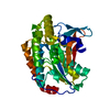
Mass: 18.015 Da / Num. of mol.: 121 / Source method: isolated from a natural source / Formula: H2O
Has protein modificationN

-
Experimental details

-
Experiment

ExperimentMethod: X-RAY DIFFRACTION / Number of used crystals: 1

-
Sample preparation

CrystalDensity Matthews: 2.63 Å3/Da / Density % sol: 53.25 %
Crystal growTemperature: 291.15 K / Method: vapor diffusion, sitting drop
Details: 0.1 M sodium acetate pH 4.5, 5% (w/v) PEG1000, 50% (v/v) ethylene glycol

-
Data collection

DiffractionMean temperature: 100 K / Serial crystal experiment: N
Diffraction sourceSource: SYNCHROTRON / Site: SSRF / Beamline: BL19U1 / Wavelength: 0.9792 Å
DetectorType: DECTRIS PILATUS 6M / Detector: PIXEL / Date: Feb 6, 2021
RadiationProtocol: SINGLE WAVELENGTH / Monochromatic (M) / Laue (L): M / Scattering type: x-ray
Radiation wavelengthWavelength: 0.9792 Å / Relative weight: 1
ReflectionResolution: 1.648→50 Å / Num. obs: 45392 / % possible obs: 100 % / Redundancy: 19 % / CC1/2: 0.998 / CC star: 1 / Rmerge(I) obs: 0.087 / Rpim(I) all: 0.021 / Rrim(I) all: 0.089 / Χ2: 1.307 / Net I/σ(I): 6.8
Reflection shell

Diffraction-ID: 1

Resolution (Å)Redundancy (%)Rmerge(I) obsNum. unique obsCC1/2CC starRpim(I) allRrim(I) allΧ2% possible all
1.65-1.6919.91.11329770.8530.960.2551.1420.446100
1.69-1.7319.50.86729810.9050.9750.2010.890.499100
1.73-1.7818.70.60330050.9410.9850.1430.620.459100
1.78-1.8319.40.46829710.9660.9910.1090.4810.492100
1.83-1.8919.50.36630120.9820.9950.0850.3750.588100
1.89-1.96200.33629760.9870.9970.0770.3450.85100
1.96-2.0319.80.1830180.9950.9990.0410.1840.612100
2.03-2.1318.50.17730230.9950.9990.0430.1820.98100
2.13-2.2419.10.11730150.9970.9990.0270.120.989100
2.24-2.3819.80.11829920.9970.9990.0270.1211.448100
2.38-2.5619.80.09330420.9980.9990.0220.0961.525100
2.56-2.8218.80.08830280.9980.9990.0210.091.977100
2.82-3.2319.10.08130560.9980.9990.0190.0832.476100
3.23-4.0717.70.07230950.99910.0180.0743.182100
4.07-50160.06632010.99910.0170.0693.49399.9

-
Processing

Software
NameVersionClassification
PHENIX(1.16_3549: ???)refinement
HKL-2000data scaling
Cootmodel building
PHENIXphasing
RefinementMethod to determine structure: AB INITIO PHASING / Resolution: 1.648→27.97 Å / SU ML: 0.21 / Cross valid method: FREE R-VALUE / σ(F): 1.34 / Phase error: 23.8 / Stereochemistry target values: ML
RfactorNum. reflection% reflection
Rfree0.2266 2258 4.98 %
Rwork0.2036 --
obs0.205 45343 99.93 %
Solvent computationShrinkage radii: 0.9 Å / VDW probe radii: 1.11 Å / Solvent model: FLAT BULK SOLVENT MODEL
Refinement stepCycle: LAST / Resolution: 1.648→27.97 Å
ProteinNucleic acidLigandSolventTotal
Num. atoms2354 0 0 121 2475
Refine LS restraints
Refine-IDTypeDev idealNumber
X-RAY DIFFRACTIONf_bond_d0.0062393
X-RAY DIFFRACTIONf_angle_d0.833233
X-RAY DIFFRACTIONf_dihedral_angle_d2.8311451
X-RAY DIFFRACTIONf_chiral_restr0.051374
X-RAY DIFFRACTIONf_plane_restr0.005414
LS refinement shell
Resolution (Å)Rfactor RfreeNum. reflection RfreeRfactor RworkNum. reflection RworkRefine-ID% reflection obs (%)
1.648-1.68390.31711230.28562662X-RAY DIFFRACTION99
1.6839-1.7230.29611430.26912660X-RAY DIFFRACTION100
1.723-1.76610.27711450.25742640X-RAY DIFFRACTION100
1.7661-1.81390.29421510.25382655X-RAY DIFFRACTION100
1.8139-1.86720.29171550.24882644X-RAY DIFFRACTION100
1.8672-1.92750.30531410.25182668X-RAY DIFFRACTION100
1.9275-1.99640.27641410.24172684X-RAY DIFFRACTION100
1.9964-2.07630.25711620.23312676X-RAY DIFFRACTION100
2.0763-2.17070.23771290.22582683X-RAY DIFFRACTION100
2.1707-2.28510.24941380.22242676X-RAY DIFFRACTION100
2.2851-2.42830.26151370.21552692X-RAY DIFFRACTION100
2.4283-2.61560.2421470.21722706X-RAY DIFFRACTION100
2.6156-2.87870.21521360.19822714X-RAY DIFFRACTION100
2.8787-3.29480.25041170.20372743X-RAY DIFFRACTION100
3.2948-4.14930.21461330.18262750X-RAY DIFFRACTION100

+
About Yorodumi

-
News

-
Feb 9, 2022. New format data for meta-information of EMDB entries

New format data for meta-information of EMDB entries

  • Version 3 of the EMDB header file is now the official format.
  • The previous official version 1.9 will be removed from the archive.

Related info.:EMDB header

External links:wwPDB to switch to version 3 of the EMDB data model

-
Aug 12, 2020. Covid-19 info

Covid-19 info

URL: https://pdbj.org/emnavi/covid19.php

New page: Covid-19 featured information page in EM Navigator.

Related info.:Covid-19 info / Mar 5, 2020. Novel coronavirus structure data

+
Mar 5, 2020. Novel coronavirus structure data

Novel coronavirus structure data

Related info.:Yorodumi Speices / Aug 12, 2020. Covid-19 info

External links:COVID-19 featured content - PDBj / Molecule of the Month (242):Coronavirus Proteases

+
Jan 31, 2019. EMDB accession codes are about to change! (news from PDBe EMDB page)

EMDB accession codes are about to change! (news from PDBe EMDB page)

  • The allocation of 4 digits for EMDB accession codes will soon come to an end. Whilst these codes will remain in use, new EMDB accession codes will include an additional digit and will expand incrementally as the available range of codes is exhausted. The current 4-digit format prefixed with “EMD-” (i.e. EMD-XXXX) will advance to a 5-digit format (i.e. EMD-XXXXX), and so on. It is currently estimated that the 4-digit codes will be depleted around Spring 2019, at which point the 5-digit format will come into force.
  • The EM Navigator/Yorodumi systems omit the EMD- prefix.

Related info.:Q: What is EMD? / ID/Accession-code notation in Yorodumi/EM Navigator

External links:EMDB Accession Codes are Changing Soon! / Contact to PDBj

+
Jul 12, 2017. Major update of PDB

Major update of PDB

  • wwPDB released updated PDB data conforming to the new PDBx/mmCIF dictionary.
  • This is a major update changing the version number from 4 to 5, and with Remediation, in which all the entries are updated.
  • In this update, many items about electron microscopy experimental information are reorganized (e.g. em_software).
  • Now, EM Navigator and Yorodumi are based on the updated data.

External links:wwPDB Remediation / Enriched Model Files Conforming to OneDep Data Standards Now Available in the PDB FTP Archive

-
Yorodumi

Thousand views of thousand structures

  • Yorodumi is a browser for structure data from EMDB, PDB, SASBDB, etc.
  • This page is also the successor to EM Navigator detail page, and also detail information page/front-end page for Omokage search.
  • The word "yorodu" (or yorozu) is an old Japanese word meaning "ten thousand". "mi" (miru) is to see.

Related info.:EMDB / PDB / SASBDB / Comparison of 3 databanks / Yorodumi Search / Aug 31, 2016. New EM Navigator & Yorodumi / Yorodumi Papers / Jmol/JSmol / Function and homology information / Changes in new EM Navigator and Yorodumi

Read more