 Movie
Movie Controller

[English] 日本語
 Yorodumi
- EMDB-51284: Structure of GPR55 in complex with G13 and synthetic agonist ML184 -
+ Open data

- Basic information
| Entry |  | |||||||||
|---|---|---|---|---|---|---|---|---|---|---|
| Title | Structure of GPR55 in complex with G13 and synthetic agonist ML184 | |||||||||
 Map data Map data | ||||||||||
 Sample Sample |
| |||||||||
 Keywords Keywords | G protein-coupled receptor / GPCR / lipid agonist / cholesterol / MEMBRANE PROTEIN | |||||||||
| Function / homology |  Function and homology information Function and homology informationD5 dopamine receptor binding / Rho-activating G protein-coupled receptor signaling pathway / regulation of fibroblast migration / cannabinoid receptor activity / Class A/1 (Rhodopsin-like receptors) / regulation of small GTPase mediated signal transduction / NRAGE signals death through JNK / branching involved in blood vessel morphogenesis / positive regulation of Rho protein signal transduction / negative regulation of vascular associated smooth muscle cell migration ...D5 dopamine receptor binding / Rho-activating G protein-coupled receptor signaling pathway / regulation of fibroblast migration / cannabinoid receptor activity / Class A/1 (Rhodopsin-like receptors) / regulation of small GTPase mediated signal transduction / NRAGE signals death through JNK / branching involved in blood vessel morphogenesis / positive regulation of Rho protein signal transduction / negative regulation of vascular associated smooth muscle cell migration / negative regulation of osteoclast differentiation / negative regulation of vascular associated smooth muscle cell proliferation / CDC42 GTPase cycle / regulation of postsynapse assembly / Rho protein signal transduction / bone resorption / RAC1 GTPase cycle / bioluminescence / guanyl-nucleotide exchange factor activity / generation of precursor metabolites and energy / brush border membrane / G protein-coupled receptor activity / adenylate cyclase-modulating G protein-coupled receptor signaling pathway / regulation of blood pressure / platelet activation / G-protein beta/gamma-subunit complex binding / Olfactory Signaling Pathway / Activation of the phototransduction cascade / G beta:gamma signalling through PLC beta / Presynaptic function of Kainate receptors / Thromboxane signalling through TP receptor / G protein-coupled acetylcholine receptor signaling pathway / adenylate cyclase-activating G protein-coupled receptor signaling pathway / G-protein activation / Activation of G protein gated Potassium channels / Inhibition of voltage gated Ca2+ channels via Gbeta/gamma subunits / Prostacyclin signalling through prostacyclin receptor / G beta:gamma signalling through CDC42 / Glucagon signaling in metabolic regulation / G beta:gamma signalling through BTK / Synthesis, secretion, and inactivation of Glucagon-like Peptide-1 (GLP-1) / ADP signalling through P2Y purinoceptor 12 / Sensory perception of sweet, bitter, and umami (glutamate) taste / photoreceptor disc membrane / Glucagon-type ligand receptors / Adrenaline,noradrenaline inhibits insulin secretion / Vasopressin regulates renal water homeostasis via Aquaporins / G alpha (z) signalling events / Glucagon-like Peptide-1 (GLP1) regulates insulin secretion / ADORA2B mediated anti-inflammatory cytokines production / cellular response to catecholamine stimulus / ADP signalling through P2Y purinoceptor 1 / G beta:gamma signalling through PI3Kgamma / adenylate cyclase-activating dopamine receptor signaling pathway / Cooperation of PDCL (PhLP1) and TRiC/CCT in G-protein beta folding / GPER1 signaling / Inactivation, recovery and regulation of the phototransduction cascade / cellular response to prostaglandin E stimulus / G-protein beta-subunit binding / heterotrimeric G-protein complex / melanosome / G alpha (12/13) signalling events / sensory perception of taste / extracellular vesicle / signaling receptor complex adaptor activity / regulation of cell shape / Thrombin signalling through proteinase activated receptors (PARs) / G protein activity / GTPase binding / Ca2+ pathway / positive regulation of cytosolic calcium ion concentration / retina development in camera-type eye / High laminar flow shear stress activates signaling by PIEZO1 and PECAM1:CDH5:KDR in endothelial cells / fibroblast proliferation / G alpha (i) signalling events / G alpha (s) signalling events / phospholipase C-activating G protein-coupled receptor signaling pathway / G alpha (q) signalling events / in utero embryonic development / Ras protein signal transduction / cell differentiation / Extra-nuclear estrogen signaling / cell population proliferation / postsynapse / positive regulation of ERK1 and ERK2 cascade / G protein-coupled receptor signaling pathway / lysosomal membrane / focal adhesion / GTPase activity / synapse / protein-containing complex binding / GTP binding / signal transduction / extracellular exosome / metal ion binding / nucleus / membrane / plasma membrane / cytosol / cytoplasm Similarity search - Function | |||||||||
| Biological species |  Homo sapiens (human) / Homo sapiens (human) /    Aequorea victoria (jellyfish) Aequorea victoria (jellyfish) | |||||||||
| Method | single particle reconstruction / cryo EM / Resolution: 2.51 Å | |||||||||
 Authors Authors | Claff T / Ebenhoch R / Weichert D | |||||||||
| Funding support | 1 items
| |||||||||
 Citation Citation | Journal: Nat Commun / Year: 2025 Title: Structural basis for lipid-mediated activation of G protein-coupled receptor GPR55. Authors: Tobias Claff / Rebecca Ebenhoch / Jörg T Kley / Aniket Magarkar / Herbert Nar / Dietmar Weichert /  Abstract: GPR55 is an orphan G protein-coupled receptor (GPCR) and represents a promising drug target for cancer, inflammation, and metabolic diseases. The endogenous activation of lipid GPCRs can be solely ...GPR55 is an orphan G protein-coupled receptor (GPCR) and represents a promising drug target for cancer, inflammation, and metabolic diseases. The endogenous activation of lipid GPCRs can be solely mediated by membrane components and different lipids have been proposed as endogenous activators of GPR55, such as cannabinoids and lysophosphatidylinositols. Here, we determine high-resolution cryo-electron microscopy structures of the activated GPR55 in complex with heterotrimeric G and two structurally diverse ligands: the putative endogenous agonist 1-palmitoyl-2-lysophosphatidylinositol (LPI) and the synthetic agonist ML184. These results reveal insights into ligand recognition at GPR55, G protein coupling and receptor activation. Notably, an orthosteric binding site opening towards the membrane is observed in both structures, enabling direct interaction of the agonists with membrane lipids. The structural observations are supported by mutagenesis and functional experiments employing G protein dissociation assays. These findings will be of importance for the structure-based development of drugs targeting GPR55. | |||||||||
| History |
|
- Structure visualization
| Supplemental images |
|---|

- Downloads & links
-EMDB archive
| Map data | emd_51284.map.gz | 25.4 MB | EMDB map data format | |
|---|---|---|---|---|
| Header (meta data) | emd-51284-v30.xmlemd-51284.xml | 20.6 KB 20.6 KB | Display Display | EMDB header |
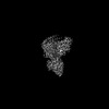
| Images |  emd_51284.png emd_51284.png | 65.4 KB | ||
| Filedesc metadata | emd-51284.cif.gz | 7.2 KB | ||
| Archive directory |  http://ftp.pdbj.org/pub/emdb/structures/EMD-51284ftp://ftp.pdbj.org/pub/emdb/structures/EMD-51284 http://ftp.pdbj.org/pub/emdb/structures/EMD-51284ftp://ftp.pdbj.org/pub/emdb/structures/EMD-51284 | HTTPS FTP |
-Validation report
| Summary document | emd_51284_validation.pdf.gz | 373.4 KB | Display | EMDB validaton report |
|---|---|---|---|---|
| Full document | emd_51284_full_validation.pdf.gz | 372.9 KB | Display | |
| Data in XML | emd_51284_validation.xml.gz | 6.9 KB | Display | |
| Data in CIF | emd_51284_validation.cif.gz | 8 KB | Display | |
| Arichive directory | https://ftp.pdbj.org/pub/emdb/validation_reports/EMD-51284ftp://ftp.pdbj.org/pub/emdb/validation_reports/EMD-51284 | HTTPS FTP |
-Related structure data
| Related structure data |  9ge2MC 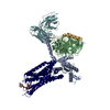 9ge3C C: citing same article ( M: atomic model generated by this map |
|---|---|
| Similar structure data |


-Links
| EMDB pages | EMDB (EBI/PDBe) / EMDataResource |
|---|---|
| Related items in Molecule of the Month |


-Map
| File | Download / File: emd_51284.map.gz / Format: CCP4 / Size: 125 MB / Type: IMAGE STORED AS FLOATING POINT NUMBER (4 BYTES) | ||||||||||||||||||||||||||||||||||||
|---|---|---|---|---|---|---|---|---|---|---|---|---|---|---|---|---|---|---|---|---|---|---|---|---|---|---|---|---|---|---|---|---|---|---|---|---|---|
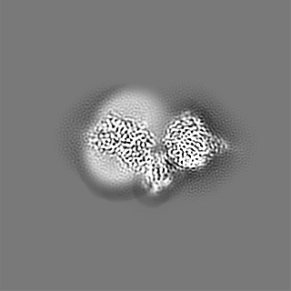
| Projections & slices | Image control
Images are generated by Spider. | ||||||||||||||||||||||||||||||||||||
| Voxel size | X=Y=Z: 0.829 Å | ||||||||||||||||||||||||||||||||||||
| Density |
| ||||||||||||||||||||||||||||||||||||
| Symmetry | Space group: 1 | ||||||||||||||||||||||||||||||||||||
| Details | EMDB XML:
|
 X (Sec.)
 Y (Row.)
 Z (Col.)


-Supplemental data
- Sample components
+Entire : Structure of GPR55 in complex with G13 and synthetic agonist ML184
+Supramolecule #1: Structure of GPR55 in complex with G13 and synthetic agonist ML184
+Supramolecule #2: Guanine nucleotide-binding proteins
+Supramolecule #3: Single-chain variable fragment ScFv16
+Supramolecule #4: G-protein coupled receptor 55 with a GFP tag
+Macromolecule #1: Guanine nucleotide-binding protein subunit alpha-13
 Trichoplusia ni (cabbage looper)
+Macromolecule #2: Guanine nucleotide-binding protein G(I)/G(S)/G(T) subunit beta-1
+Macromolecule #3: Single-chain variable fragment ScFv16
+Macromolecule #4: Guanine nucleotide-binding protein G(I)/G(S)/G(O) subunit gamma-2
+Macromolecule #5: Green fluorescent protein,G-protein coupled receptor 55
+Macromolecule #7: 3-[4-(2,3-dimethylphenyl)piperazin-1-yl]carbonyl-N,N-dimethyl-4-p...
+Macromolecule #8: CHOLESTEROL

+Macromolecule #9: water

-Experimental details
-Structure determination
| Method | cryo EM |
|---|---|
 Processing Processing | single particle reconstruction |
| Aggregation state | particle |
-Sample preparation
| Buffer | pH: 7.5 |
|---|---|
| Vitrification | Cryogen name: ETHANE |
- Electron microscopy
| Microscope | FEI TITAN KRIOS |
|---|---|
| Image recording | Film or detector model: GATAN K3 (6k x 4k) / Average electron dose: 45.0 e/Å2 |
| Electron beam | Acceleration voltage: 300 kV / Electron source:  FIELD EMISSION GUN FIELD EMISSION GUN |
| Electron optics | Illumination mode: OTHER / Imaging mode: BRIGHT FIELD / Nominal defocus max: 3.0 µm / Nominal defocus min: 0.8 µm / Nominal magnification: 105000 |
| Experimental equipment |  Model: Titan Krios / Image courtesy: FEI Company |
