 Movie
Movie Controller

[English] 日本語
 Yorodumi
- EMDB-50562: Aerolysin heptamer in membrane inserted form reconstituted in amp... -
+ Open data

- Basic information
| Entry | 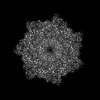 | ||||||||||||
|---|---|---|---|---|---|---|---|---|---|---|---|---|---|
| Title | Aerolysin heptamer in membrane inserted form reconstituted in amphipoles. | ||||||||||||
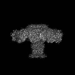
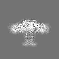
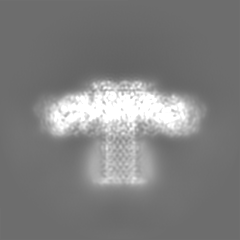
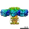
 Map data Map data | postprocessed map of aerolysin heptamer in A8-35. | ||||||||||||
 Sample Sample |
| ||||||||||||
 Keywords Keywords | pore forming toxin / aerolysin / amphipole reconstitution / cryo-EM / TOXIN | ||||||||||||
| Function / homology |  Function and homology information Function and homology informationsymbiont-mediated cytolysis of host cell / toxin activity / host cell plasma membrane / extracellular region / identical protein binding Similarity search - Function | ||||||||||||
| Biological species |  Aeromonas hydrophila (bacteria) Aeromonas hydrophila (bacteria) | ||||||||||||
| Method | single particle reconstruction / cryo EM / Resolution: 2.2 Å | ||||||||||||
 Authors Authors | Iacovache I / Zuber B | ||||||||||||
| Funding support |  Switzerland, 3 items Switzerland, 3 items
| ||||||||||||
 Citation Citation | Journal: J Am Chem Soc / Year: 2025 Title: Aerolysin Nanopore Structures Revealed at High Resolution in a Lipid Environment. Authors: Jana S Anton / Ioan Iacovache / Juan F Bada Juarez / Luciano A Abriata / Louis W Perrin / Chan Cao / Maria J Marcaida / Benoît Zuber / Matteo Dal Peraro / Abstract: Aerolysin is a β-pore-forming toxin produced by most Aeromonas bacteria, which has attracted large attention in the field of nanopore sensing due to its narrow and charged pore lumen. Structurally ...Aerolysin is a β-pore-forming toxin produced by most Aeromonas bacteria, which has attracted large attention in the field of nanopore sensing due to its narrow and charged pore lumen. Structurally similar proteins, belonging to the aerolysin-like family, are present throughout all kingdoms of life, but very few of them have been structurally characterized in a lipid environment. Here, we present the first high-resolution atomic cryo-EM structures of aerolysin prepore and pore in a membrane-like environment. These structures allow the identification of key interactions, which are relevant for understanding the pore formation mechanism and for correctly positioning the pore β-barrel and its anchoring β-turn motif in the membrane. Moreover, we elucidate at high resolution the architecture of key pore mutations and precisely identify four constriction rings in the pore lumen that are highly relevant for nanopore sensing experiments. | ||||||||||||
| History |
|
- Structure visualization
| Supplemental images |
|---|

- Downloads & links
-EMDB archive
| Map data | emd_50562.map.gz | 7 MB | EMDB map data format | |
|---|---|---|---|---|
| Header (meta data) | emd-50562-v30.xmlemd-50562.xml | 19.7 KB 19.7 KB | Display Display | EMDB header |
| FSC (resolution estimation) | emd_50562_fsc.xml | 8.5 KB | Display | FSC data file |
| Images |  emd_50562.png emd_50562.png | 81.8 KB | ||
| Masks | emd_50562_msk_1.map | 52.7 MB | Mask map | |
| Filedesc metadata | emd-50562.cif.gz | 6.5 KB | ||
| Others | emd_50562_additional_1.map.gzemd_50562_half_map_1.map.gzemd_50562_half_map_2.map.gz | 40.5 MB 40.7 MB 40.7 MB | ||
| Archive directory |  http://ftp.pdbj.org/pub/emdb/structures/EMD-50562ftp://ftp.pdbj.org/pub/emdb/structures/EMD-50562 http://ftp.pdbj.org/pub/emdb/structures/EMD-50562ftp://ftp.pdbj.org/pub/emdb/structures/EMD-50562 | HTTPS FTP |
-Related structure data
| Related structure data |  9fmlMC  9fm6C  9fmxC  9fnpC  9fnqC M: atomic model generated by this map C: citing same article ( |
|---|---|
| Similar structure data |


-Links
| EMDB pages | EMDB (EBI/PDBe) / EMDataResource |
|---|---|
| Related items in Molecule of the Month |


-Map
| File | Download / File: emd_50562.map.gz / Format: CCP4 / Size: 52.7 MB / Type: IMAGE STORED AS FLOATING POINT NUMBER (4 BYTES) | ||||||||||||||||||||||||||||||||||||
|---|---|---|---|---|---|---|---|---|---|---|---|---|---|---|---|---|---|---|---|---|---|---|---|---|---|---|---|---|---|---|---|---|---|---|---|---|---|
| Annotation | postprocessed map of aerolysin heptamer in A8-35. | ||||||||||||||||||||||||||||||||||||
| Projections & slices | Image control
Images are generated by Spider. | ||||||||||||||||||||||||||||||||||||
| Voxel size | X=Y=Z: 0.94 Å | ||||||||||||||||||||||||||||||||||||
| Density |
| ||||||||||||||||||||||||||||||||||||
| Symmetry | Space group: 1 | ||||||||||||||||||||||||||||||||||||
| Details | EMDB XML:
|
 Z (Sec.)
 Y (Row.)
 X (Col.)


-Supplemental data
-Mask #1
| File | emd_50562_msk_1.map | ||||||||||||
|---|---|---|---|---|---|---|---|---|---|---|---|---|---|
| Projections & Slices |
| ||||||||||||
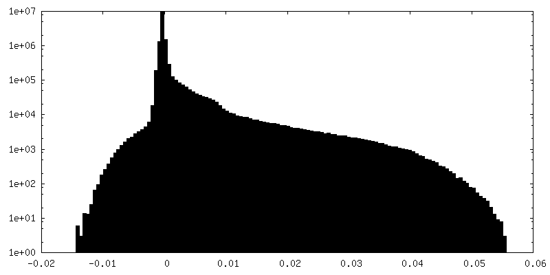
| Density Histograms |


-Additional map: refinement map
| File | emd_50562_additional_1.map | ||||||||||||
|---|---|---|---|---|---|---|---|---|---|---|---|---|---|
| Annotation | refinement map | ||||||||||||
| Projections & Slices |
| ||||||||||||
| Density Histograms |


-Half map: half map 2
| File | emd_50562_half_map_1.map | ||||||||||||
|---|---|---|---|---|---|---|---|---|---|---|---|---|---|
| Annotation | half map 2 | ||||||||||||
| Projections & Slices |
| ||||||||||||
| Density Histograms |


-Half map: half map 1
| File | emd_50562_half_map_2.map | ||||||||||||
|---|---|---|---|---|---|---|---|---|---|---|---|---|---|
| Annotation | half map 1 | ||||||||||||
| Projections & Slices |
| ||||||||||||
| Density Histograms |


- Sample components
-Entire : aerolysin heptamer in A8-35
| Entire | Name: aerolysin heptamer in A8-35 |
|---|---|
| Components |
|
-Supramolecule #1: aerolysin heptamer in A8-35
| Supramolecule | Name: aerolysin heptamer in A8-35 / type: complex / ID: 1 / Parent: 0 / Macromolecule list: all Details: Heptamerization was induced by trypsinization of proaerolysin. |
|---|---|
| Source (natural) | Organism: Aeromonas hydrophila (bacteria) |
| Molecular weight | Theoretical: 350 KDa |
-Macromolecule #1: Aerolysin
| Macromolecule | Name: Aerolysin / type: protein_or_peptide / ID: 1 / Number of copies: 7 / Enantiomer: LEVO |
|---|---|
| Source (natural) | Organism: Aeromonas hydrophila (bacteria) |
| Molecular weight | Theoretical: 51.980453 KDa |
| Recombinant expression | Organism: |
| Sequence | String: AEPVYPDQLR LFSLGQGVCG DKYRPVNREE AQSVKSNIVG MMGQWQISGL ANGWVIMGPG YNGEIKPGTA SNTWCYPTNP VTGEIPTLS ALDIPDGDEV DVQWRLVHDS ANFIKPTSYL AHYLGYAWVG GNHSQYVGED MDVTRDGDGW VIRGNNDGGC D GYRCGDKT ...String: AEPVYPDQLR LFSLGQGVCG DKYRPVNREE AQSVKSNIVG MMGQWQISGL ANGWVIMGPG YNGEIKPGTA SNTWCYPTNP VTGEIPTLS ALDIPDGDEV DVQWRLVHDS ANFIKPTSYL AHYLGYAWVG GNHSQYVGED MDVTRDGDGW VIRGNNDGGC D GYRCGDKT AIKVSNFAYN LDPDSFKHGD VTQSDRQLVK TVVGWAVNDS DTPQSGYDVT LRYDTATNWS KTNTYGLSEK VT TKNKFKW PLVGETELSI EIAANQSWAS QNGGSTTTSL SQSVRPTVPA RSKIPVKIEL YKADISYPYE FKADVSYDLT LSG FLRWGG NAWYTHPDNR PNWNHTFVIG PYKDKASSIR YQWDKRYIPG EVKWWDWNWT IQQNGLSTMQ NNLARVLRPV RAGI TGDFS AESQFAGNIE IGAPVPLAAD SKVRRARSVD GAGQGLRLEI PLDAQELSGL GFNNVSLSVT PAANQ UniProtKB: Aerolysin |
-Experimental details
-Structure determination
| Method | cryo EM |
|---|---|
 Processing Processing | single particle reconstruction |
| Aggregation state | particle |
-Sample preparation
| Concentration | 1 mg/mL |
|---|---|
| Buffer | pH: 7.4 / Details: 20mM Hepes 7.4 100mM NaCl 0.05% A8-35 |
| Grid | Model: Quantifoil R2/1 / Material: COPPER / Mesh: 200 / Support film - Material: CARBON / Support film - topology: CONTINUOUS / Support film - Film thickness: 2 / Pretreatment - Type: GLOW DISCHARGE / Pretreatment - Time: 5 sec. / Pretreatment - Atmosphere: AIR / Pretreatment - Pressure: 2e-05 kPa |
| Vitrification | Cryogen name: ETHANE / Chamber humidity: 100 % / Chamber temperature: 298 K / Instrument: FEI VITROBOT MARK IV |
- Electron microscopy
| Microscope | FEI TITAN KRIOS |
|---|---|
| Specialist optics | Energy filter - Name: TFS Selectris / Energy filter - Slit width: 20 eV |
| Image recording | Film or detector model: FEI FALCON IV (4k x 4k) / Average electron dose: 40.0 e/Å2 |
| Electron beam | Acceleration voltage: 300 kV / Electron source:  FIELD EMISSION GUN FIELD EMISSION GUN |
| Electron optics | Illumination mode: FLOOD BEAM / Imaging mode: BRIGHT FIELD / Cs: 2.7 mm / Nominal defocus max: 3.0 µm / Nominal defocus min: 1.2 µm |
| Sample stage | Specimen holder model: FEI TITAN KRIOS AUTOGRID HOLDER / Cooling holder cryogen: NITROGEN |
| Experimental equipment |  Model: Titan Krios / Image courtesy: FEI Company |
+Image processing

-Atomic model buiding 1
| Initial model | Chain - Source name: Other / Chain - Initial model type: other / Details: modelangelo |
|---|---|
| Details | ModelAngelo was used with the final map to generate the first model. |
| Refinement | Protocol: OTHER |
| Output model | PDB-9fml: |
