 Movie
Movie Controller


+ Open data

- Basic information
| Entry |  | |||||||||
|---|---|---|---|---|---|---|---|---|---|---|
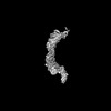
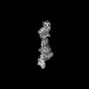
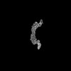
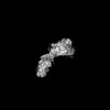
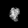
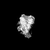
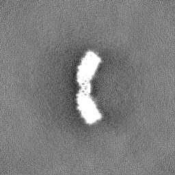
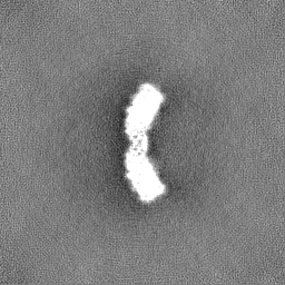
| Title | P116 (amino acids 246-818) dimer in the full state | |||||||||
 Map data Map data | ||||||||||
 Sample Sample |
| |||||||||
 Keywords Keywords | Lipid Transfer Protein / Mycoplasma pneumoniae / LIPID BINDING PROTEIN | |||||||||
| Function / homology | : / : / P116-like protein / membrane / Uncharacterized protein MG075 homolog Function and homology information Function and homology information | |||||||||
| Biological species |  Mycoplasmoides pneumoniae M129 (bacteria) Mycoplasmoides pneumoniae M129 (bacteria) | |||||||||
| Method | single particle reconstruction / cryo EM / Resolution: 6.52 Å | |||||||||
 Authors Authors | Mager S | |||||||||
| Funding support |  Germany, 1 items Germany, 1 items
| |||||||||
 Citation Citation | Journal: Nat Struct Mol Biol / Year: 2023 Title: Essential protein P116 extracts cholesterol and other indispensable lipids for Mycoplasmas. Authors: Lasse Sprankel / David Vizarraga / Jesús Martín / Sina Manger / Jakob Meier-Credo / Marina Marcos / Josep Julve / Noemi Rotllan / Margot P Scheffer / Joan Carles Escolà-Gil / Julian D ...Authors: Lasse Sprankel / David Vizarraga / Jesús Martín / Sina Manger / Jakob Meier-Credo / Marina Marcos / Josep Julve / Noemi Rotllan / Margot P Scheffer / Joan Carles Escolà-Gil / Julian D Langer / Jaume Piñol / Ignacio Fita / Achilleas S Frangakis /  Abstract: Mycoplasma pneumoniae, responsible for approximately 30% of community-acquired human pneumonia, needs to extract lipids from the host environment for survival and proliferation. Here, we report a ...Mycoplasma pneumoniae, responsible for approximately 30% of community-acquired human pneumonia, needs to extract lipids from the host environment for survival and proliferation. Here, we report a comprehensive structural and functional analysis of the previously uncharacterized protein P116 (MPN_213). Single-particle cryo-electron microscopy of P116 reveals a homodimer presenting a previously unseen fold, forming a huge hydrophobic cavity, which is fully accessible to solvent. Lipidomics analysis shows that P116 specifically extracts lipids such as phosphatidylcholine, sphingomyelin and cholesterol. Structures of different conformational states reveal the mechanism by which lipids are extracted. This finding immediately suggests a way to control Mycoplasma infection by interfering with lipid uptake. | |||||||||
| History |
|
- Structure visualization
| Supplemental images |
|---|

- Downloads & links
-EMDB archive
| Map data | emd_50314.map.gz | 32.4 MB | EMDB map data format | |
|---|---|---|---|---|
| Header (meta data) | emd-50314-v30.xmlemd-50314.xml | 21.3 KB 21.3 KB | Display Display | EMDB header |
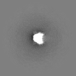
| Images |  emd_50314.png emd_50314.png | 52.6 KB | ||
| Masks | emd_50314_msk_1.map | 64 MB | Mask map | |
| Filedesc metadata | emd-50314.cif.gz | 6.5 KB | ||
| Others | emd_50314_half_map_1.map.gzemd_50314_half_map_2.map.gz | 59.4 MB 59.4 MB | ||
| Archive directory |  http://ftp.pdbj.org/pub/emdb/structures/EMD-50314ftp://ftp.pdbj.org/pub/emdb/structures/EMD-50314 http://ftp.pdbj.org/pub/emdb/structures/EMD-50314ftp://ftp.pdbj.org/pub/emdb/structures/EMD-50314 | HTTPS FTP |
-Related structure data
| Related structure data |  9fchMC M: atomic model generated by this map C: citing same article ( |
|---|---|
| Similar structure data |

-Links
| EMDB pages | EMDB (EBI/PDBe) / EMDataResource |
|---|
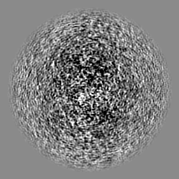
-Map
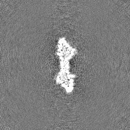
| File | Download / File: emd_50314.map.gz / Format: CCP4 / Size: 64 MB / Type: IMAGE STORED AS FLOATING POINT NUMBER (4 BYTES) | ||||||||||||||||||||||||||||||||||||
|---|---|---|---|---|---|---|---|---|---|---|---|---|---|---|---|---|---|---|---|---|---|---|---|---|---|---|---|---|---|---|---|---|---|---|---|---|---|
| Projections & slices | Image control
Images are generated by Spider. | ||||||||||||||||||||||||||||||||||||
| Voxel size | X=Y=Z: 1.638 Å | ||||||||||||||||||||||||||||||||||||
| Density |
| ||||||||||||||||||||||||||||||||||||
| Symmetry | Space group: 1 | ||||||||||||||||||||||||||||||||||||
| Details | EMDB XML:
|
 Z (Sec.)
 Y (Row.)
 X (Col.)


-Supplemental data
-Mask #1
| File | emd_50314_msk_1.map | ||||||||||||
|---|---|---|---|---|---|---|---|---|---|---|---|---|---|
| Projections & Slices |
| ||||||||||||
| Density Histograms |


-Half map: #1
| File | emd_50314_half_map_1.map | ||||||||||||
|---|---|---|---|---|---|---|---|---|---|---|---|---|---|
| Projections & Slices |
| ||||||||||||
| Density Histograms |


-Half map: #2
| File | emd_50314_half_map_2.map | ||||||||||||
|---|---|---|---|---|---|---|---|---|---|---|---|---|---|
| Projections & Slices |
| ||||||||||||
| Density Histograms |


- Sample components
-Entire : P116 from Mycoplasma pneumoniae (aa 246-818; full state)
| Entire | Name: P116 from Mycoplasma pneumoniae (aa 246-818; full state) |
|---|---|
| Components |
|
-Supramolecule #1: P116 from Mycoplasma pneumoniae (aa 246-818; full state)
| Supramolecule | Name: P116 from Mycoplasma pneumoniae (aa 246-818; full state) type: complex / ID: 1 / Parent: 0 / Macromolecule list: all |
|---|---|
| Source (natural) | Organism: Mycoplasmoides pneumoniae M129 (bacteria) |
-Macromolecule #1: Uncharacterized protein MG075 homolog
| Macromolecule | Name: Uncharacterized protein MG075 homolog / type: protein_or_peptide / ID: 1 / Number of copies: 2 / Enantiomer: LEVO |
|---|---|
| Source (natural) | Organism: Mycoplasmoides pneumoniae M129 (bacteria) |
| Molecular weight | Theoretical: 64.38675 KDa |
| Recombinant expression | Organism: |
| Sequence | String: GVDVFEAQKN LVGKGKYLNT HVKAEDVKKD VNANIKNQFD IAKIIAELMG KALKEFGNQQ EGQPLSFLKV MDKVKEDFEK LFNLVRPGL GKFVKDLIQS SSQAENKITV YKLIFDNKKT ILNLLKELSI PELNSSLGLV DVLFDGITDS DGLYERLQSF K DLIVPAVK ...String: GVDVFEAQKN LVGKGKYLNT HVKAEDVKKD VNANIKNQFD IAKIIAELMG KALKEFGNQQ EGQPLSFLKV MDKVKEDFEK LFNLVRPGL GKFVKDLIQS SSQAENKITV YKLIFDNKKT ILNLLKELSI PELNSSLGLV DVLFDGITDS DGLYERLQSF K DLIVPAVK TNEKTAALSP LIEELLTQKD TYVFDLIQKH KGILTNLLKN FLADFQKSTP FMADQVAIFT ELFDNEGAFD LF GEADFVD KIAELFLTKR TVKNGEKIET KDSLLVTSLK SLLGEKVAAL GDLLDSYIFK NELLNRSVEV AKAEAKDTKG ATD YKKEQA KALKKLFKHI GENTLSKTNL DKITLKEVKN TENVELEETE TTLKVKKLDV EYKVELGNFE IKNGLIKAML EFLP DTKDL ETTLDKLLFK GESYKAMKDK YIKEGFPGYG WAKGVVPGAF ESIENTFKSA IDKTKSIRDL FGDMLFGNDL SSVKE TDSF ITLGGSFDIK YGGENLNVLP AYYSLINSEI GYQIIGVDTT IDATKVKVEL KNKEYKGKSP AINGQVKLSQ SFFNVW TNM FDSITKQIFQ UniProtKB: Uncharacterized protein MG075 homolog |
-Experimental details
-Structure determination
| Method | cryo EM |
|---|---|
 Processing Processing | single particle reconstruction |
| Aggregation state | particle |
-Sample preparation
| Concentration | 3 mg/mL |
|---|---|
| Buffer | pH: 7.4 / Details: 20 mM Tris-HCl |
| Grid | Model: C-flat-1.2/1.3 |
| Vitrification | Cryogen name: ETHANE / Chamber humidity: 100 % / Chamber temperature: 4 K / Instrument: FEI VITROBOT MARK IV |
- Electron microscopy
| Microscope | FEI TITAN KRIOS |
|---|---|
| Image recording | Film or detector model: GATAN K3 BIOQUANTUM (6k x 4k) / Average electron dose: 50.0 e/Å2 |
| Electron beam | Acceleration voltage: 300 kV / Electron source:  FIELD EMISSION GUN FIELD EMISSION GUN |
| Electron optics | C2 aperture diameter: 70.0 µm / Illumination mode: FLOOD BEAM / Imaging mode: BRIGHT FIELD / Cs: 2.7 mm / Nominal defocus max: 3.5 µm / Nominal defocus min: 0.8 µm |
| Experimental equipment |  Model: Titan Krios / Image courtesy: FEI Company |
