[English] 日本語
 Yorodumi
- EMDB-50185: KS + AT di-domain of polyketide synthase 13 in Mycobacterium tube... -
+ Open data
- Basic information
| Entry |  | |||||||||
|---|---|---|---|---|---|---|---|---|---|---|
| Title | KS + AT di-domain of polyketide synthase 13 in Mycobacterium tuberculosis | |||||||||
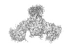
 Map data Map data | Final map for Pks13 from M. tuberculosis | |||||||||
 Sample Sample |
| |||||||||
 Keywords Keywords | Mycolic acid synthesis / Claisen condensation / cell wall synthesis / BIOSYNTHETIC PROTEIN | |||||||||
| Function / homology |  Function and homology information Function and homology informationDIM/DIP cell wall layer assembly / fatty acid synthase activity / phosphopantetheine binding / 3-oxoacyl-[acyl-carrier-protein] synthase activity / Transferases; Acyltransferases; Transferring groups other than aminoacyl groups / fatty acid biosynthetic process / cytoplasm Similarity search - Function | |||||||||
| Biological species |   Mycobacterium tuberculosis H37Rv (bacteria) Mycobacterium tuberculosis H37Rv (bacteria) | |||||||||
| Method | single particle reconstruction / cryo EM / Resolution: 3.4 Å | |||||||||
 Authors Authors | Johnston HE / Futterer K | |||||||||
| Funding support |  United Kingdom, 2 items United Kingdom, 2 items
| |||||||||
 Citation Citation |  Journal: Microbiology (Reading) / Year: 2024 Journal: Microbiology (Reading) / Year: 2024Title: Cryo-electron microscopy structure of the di-domain core of polyketide synthase 13, essential for mycobacterial mycolic acid synthesis. Authors: Hannah E Johnston / Sarah M Batt / Alistair K Brown / Christos G Savva / Gurdyal S Besra / Klaus Fütterer /  Abstract: Mycobacteria are known for their complex cell wall, which comprises layers of peptidoglycan, polysaccharides and unusual fatty acids known as mycolic acids that form their unique outer membrane. ...Mycobacteria are known for their complex cell wall, which comprises layers of peptidoglycan, polysaccharides and unusual fatty acids known as mycolic acids that form their unique outer membrane. Polyketide synthase 13 (Pks13) of , the bacterial organism causing tuberculosis, catalyses the last step of mycolic acid synthesis prior to export to and assembly in the cell wall. Due to its essentiality, Pks13 is a target for several novel anti-tubercular inhibitors, but its 3D structure and catalytic reaction mechanism remain to be fully elucidated. Here, we report the molecular structure of the catalytic core domains of Pks13 (Mt-Pks13), determined by transmission cryo-electron microscopy (cryoEM) to a resolution of 3.4 Å. We observed a homodimeric assembly comprising the ketoacyl synthase (KS) domain at the centre, mediating dimerization, and the acyltransferase (AT) domains protruding in opposite directions from the central KS domain dimer. In addition to the KS-AT di-domains, the cryoEM map includes features not covered by the di-domain structural model that we predicted to contain a dimeric domain similar to dehydratases, yet likely lacking catalytic function. Analytical ultracentrifugation data indicate a pH-dependent equilibrium between monomeric and dimeric assembly states, while comparison with the previously determined structures of Pks13 indicates architectural flexibility. Combining the experimentally determined structure with modelling in AlphaFold2 suggests a structural scaffold with a relatively stable dimeric core, which combines with considerable conformational flexibility to facilitate the successive steps of the Claisen-type condensation reaction catalysed by Pks13. | |||||||||
| History |
|
- Structure visualization
| Supplemental images |
|---|
- Downloads & links
-EMDB archive
| Map data |  emd_50185.map.gz emd_50185.map.gz | 10.2 MB |  EMDB map data format EMDB map data format | |
|---|---|---|---|---|
| Header (meta data) |  emd-50185-v30.xml emd-50185-v30.xml emd-50185.xml emd-50185.xml | 17.1 KB 17.1 KB | Display Display |  EMDB header EMDB header |
| Images |  emd_50185.png emd_50185.png | 71.1 KB | ||
| Filedesc metadata |  emd-50185.cif.gz emd-50185.cif.gz | 6.8 KB | ||
| Others |  emd_50185_half_map_1.map.gz emd_50185_half_map_1.map.gz emd_50185_half_map_2.map.gz emd_50185_half_map_2.map.gz | 141.7 MB 141.5 MB | ||
| Archive directory |  http://ftp.pdbj.org/pub/emdb/structures/EMD-50185 http://ftp.pdbj.org/pub/emdb/structures/EMD-50185 ftp://ftp.pdbj.org/pub/emdb/structures/EMD-50185 ftp://ftp.pdbj.org/pub/emdb/structures/EMD-50185 | HTTPS FTP |
-Validation report
| Summary document |  emd_50185_validation.pdf.gz emd_50185_validation.pdf.gz | 858 KB | Display |  EMDB validaton report EMDB validaton report |
|---|---|---|---|---|
| Full document |  emd_50185_full_validation.pdf.gz emd_50185_full_validation.pdf.gz | 857.6 KB | Display | |
| Data in XML |  emd_50185_validation.xml.gz emd_50185_validation.xml.gz | 14.9 KB | Display | |
| Data in CIF |  emd_50185_validation.cif.gz emd_50185_validation.cif.gz | 17.8 KB | Display | |
| Arichive directory |  https://ftp.pdbj.org/pub/emdb/validation_reports/EMD-50185 https://ftp.pdbj.org/pub/emdb/validation_reports/EMD-50185 ftp://ftp.pdbj.org/pub/emdb/validation_reports/EMD-50185 ftp://ftp.pdbj.org/pub/emdb/validation_reports/EMD-50185 | HTTPS FTP |
-Related structure data
| Related structure data |  9f48MC M: atomic model generated by this map C: citing same article ( |
|---|---|
| Similar structure data | Similarity search - Function & homology  F&H Search F&H Search |
- Links
Links
| EMDB pages |  EMDB (EBI/PDBe) / EMDB (EBI/PDBe) /  EMDataResource EMDataResource |
|---|---|
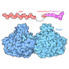
| Related items in Molecule of the Month |
- Map
Map
| File |  Download / File: emd_50185.map.gz / Format: CCP4 / Size: 178 MB / Type: IMAGE STORED AS FLOATING POINT NUMBER (4 BYTES) Download / File: emd_50185.map.gz / Format: CCP4 / Size: 178 MB / Type: IMAGE STORED AS FLOATING POINT NUMBER (4 BYTES) | ||||||||||||||||||||||||||||||||||||
|---|---|---|---|---|---|---|---|---|---|---|---|---|---|---|---|---|---|---|---|---|---|---|---|---|---|---|---|---|---|---|---|---|---|---|---|---|---|
| Annotation | Final map for Pks13 from M. tuberculosis | ||||||||||||||||||||||||||||||||||||
| Projections & slices | Image control
Images are generated by Spider. | ||||||||||||||||||||||||||||||||||||
| Voxel size | X=Y=Z: 1.09 Å | ||||||||||||||||||||||||||||||||||||
| Density |
| ||||||||||||||||||||||||||||||||||||
| Symmetry | Space group: 1 | ||||||||||||||||||||||||||||||||||||
| Details | EMDB XML:
|
-Supplemental data
-Half map: Half map 1
| File | emd_50185_half_map_1.map | ||||||||||||
|---|---|---|---|---|---|---|---|---|---|---|---|---|---|
| Annotation | Half map 1 | ||||||||||||
| Projections & Slices |
| ||||||||||||
| Density Histograms |
-Half map: Half map 2
| File | emd_50185_half_map_2.map | ||||||||||||
|---|---|---|---|---|---|---|---|---|---|---|---|---|---|
| Annotation | Half map 2 | ||||||||||||
| Projections & Slices |
| ||||||||||||
| Density Histograms |
- Sample components
-Entire : Subunit of Pks13
| Entire | Name: Subunit of Pks13 |
|---|---|
| Components |
|
-Supramolecule #1: Subunit of Pks13
| Supramolecule | Name: Subunit of Pks13 / type: complex / ID: 1 / Parent: 0 / Macromolecule list: all |
|---|---|
| Source (natural) | Organism:  |
| Molecular weight | Theoretical: 187 KDa |
-Macromolecule #1: Polyketide synthase Pks13
| Macromolecule | Name: Polyketide synthase Pks13 / type: protein_or_peptide / ID: 1 / Number of copies: 2 / Enantiomer: LEVO EC number: Transferases; Acyltransferases; Transferring groups other than aminoacyl groups |
|---|---|
| Source (natural) | Organism:  Mycobacterium tuberculosis H37Rv (bacteria) Mycobacterium tuberculosis H37Rv (bacteria) |
| Molecular weight | Theoretical: 186.642188 KDa |
| Recombinant expression | Organism:  |
| Sequence | String: MADVAESQEN APAERAELTV PEMRQWLRNW VGKAVGKAPD SIDESVPMVE LGLSSRDAVA MAADIEDLTG VTLSVAVAFA HPTIESLAT RIIEGEPETD LAGDDAEDWS RTGPAERVDI AIVGLSTRFP GEMNTPEQTW QALLEGRDGI TDLPDGRWSE F LEEPRLAA ...String: MADVAESQEN APAERAELTV PEMRQWLRNW VGKAVGKAPD SIDESVPMVE LGLSSRDAVA MAADIEDLTG VTLSVAVAFA HPTIESLAT RIIEGEPETD LAGDDAEDWS RTGPAERVDI AIVGLSTRFP GEMNTPEQTW QALLEGRDGI TDLPDGRWSE F LEEPRLAA RVAGARTRGG YLKDIKGFDS EFFAVAKTEA DNIDPQQRMA LELTWEALEH ARIPASSLRG QAVGVYIGSS TN DYSFLAV SDPTVAHPYA ITGTSSSIIA NRVSYFYDFH GPSVTIDTAC SSSLVAIHQG VQALRNGEAD VVVAGGVNAL ITP MVTLGF DEIGAVLAPD GRIKSFSADA DGYTRSEGGG MLVLKRVDDA RRDGDAILAV IAGSAVNHDG RSNGLIAPNQ DAQA DVLRR AYKDAGIDPR TVDYIEAHGT GTILGDPIEA EALGRVVGRG RPADRPALLG AVKTNVGHLE SAAGAASMAK VVLAL QHDK LPPSINFAGP SPYIDFDAMR LKMITTPTDW PRYGGYALAG VSSFGFGGAN AHVVVREVLP RDVVEKEPEP EPEPKA AAE PAEAPTLAGH ALRFDEFGNI ITDSAVAEEP EPELPGVTEE ALRLKEAALE ELAAQEVTAP LVPLAVSAFL TSRKKAA AA ELADWMQSPE GQASSLESIG RSLSRRNHGR SRAVVLAHDH DEAIKGLRAV AAGKQAPNVF SVDGPVTTGP VWVLAGFG A QHRKMGKSLY LRNEVFAAWI EKVDALVQDE LGYSVLELIL DDAQDYGIET TQVTIFAIQI ALGELLRHHG AKPAAVIGQ SLGEAASAYF AGGLSLRDAT RAICSRSHLM GEGEAMLFGE YIRLMALVEY SADEIREVFS DFPDLEVCVY AAPTQTVIGG PPEQVDAIL ARAEAEGKFA RKFATKGASH TSQMDPLLGE LTAELQGIKP TSPTCGIFST VHEGRYIKPG GEPIHDVEYW K KGLRHSVY FTHGIRNAVD SGHTTFLELA PNPVALMQVA LTTADAGLHD AQLIPTLARK QDEVSSMVST MAQLYVYGHD LD IRTLFSR ASGPQDYANI PPTRFKRKEH WLPAHFSGDG STYMPGTHVA LPDGRHVWEY APRDGNVDLA ALVRAAAAHV LPD AQLTAA EQRAVPGDGA RLVTTMTRHP GGASVQVHAR IDESFTLVYD ALVSRAGSES VLPTAVGAAT AIAVADGAPV APET PAEDA DAETLSDSLT TRYMPSGMTR WSPDSGETIA ERLGLIVGSA MGYEPEDLPW EVPLIELGLD SLMAVRIKNR VEYDF DLPP IQLTAVRDAN LYNVEKLIEY AVEHRDEVQQ LHEHQKTQTA EEIARAQAEL LHGKVGKTEP VDSEAGVALP SPQNGE QPN PTGPALNVDV PPRDAAERVT FATWAIVTGK SPGGIFNELP RLDDEAAAKI AQRLSERAEG PITAEDVLTS SNIEALA DK VRTYLEAGQI DGFVRTLRAR PEAGGKVPVF VFHPAGGSTV VYEPLLGRLP ADTPMYGFER VEGSIEERAQ QYVPKLIE M QGDGPYVLVG WSLGGVLAYA CAIGLRRLGK DVRFVGLIDA VRAGEEIPQT KEEIRKRWDR YAAFAEKTFN VTIPAIPYE QLEELDDEGQ VRFVLDAVSQ SGVQIPAGII EHQRTSYLDN RAIDTAQIQP YDGHVTLYMA DRYHDDAIMF EPRYAVRQPD GGWGEYVSD LEVVPIGGEH IQAIDEPIIA KVGEHMSRAL GQIEADRTSE VGKQ UniProtKB: Polyketide synthase Pks13 |
-Experimental details
-Structure determination
| Method | cryo EM |
|---|---|
 Processing Processing | single particle reconstruction |
| Aggregation state | particle |
- Sample preparation
| Concentration | 0.9 mg/mL |
|---|---|
| Buffer | pH: 7.9 / Component - Concentration: 20.0 mM / Component - Formula: C4H11NO3 / Component - Name: Tris Details: 20 mM Tris-HCl pH 7.9, 50 mM NaCl, 2.5 mM beta-mercaptoethanol |
| Grid | Model: Quantifoil R1.2/1.3 / Material: GOLD / Support film - Material: CARBON / Support film - topology: HOLEY / Pretreatment - Type: GLOW DISCHARGE / Pretreatment - Time: 60 sec. |
| Vitrification | Cryogen name: ETHANE / Chamber humidity: 100 % / Chamber temperature: 291 K / Instrument: FEI VITROBOT MARK IV |
- Electron microscopy
| Microscope | FEI TITAN KRIOS |
|---|---|
| Image recording | Film or detector model: GATAN K3 (6k x 4k) / Number grids imaged: 1 / Number real images: 1412 / Average exposure time: 3.0 sec. / Average electron dose: 16.5 e/Å2 / Details: movie mode |
| Electron beam | Acceleration voltage: 300 kV / Electron source:  FIELD EMISSION GUN FIELD EMISSION GUN |
| Electron optics | C2 aperture diameter: 100.0 µm / Illumination mode: FLOOD BEAM / Imaging mode: OTHER / Cs: 2.7 mm / Nominal defocus max: 2.7 µm / Nominal defocus min: 1.5 µm |
| Sample stage | Specimen holder model: FEI TITAN KRIOS AUTOGRID HOLDER |
| Experimental equipment |  Model: Titan Krios / Image courtesy: FEI Company |
 Movie
Movie Controller


 Z (Sec.)
 Y (Row.)
 X (Col.)


