Movie
Movie Controller

[English] 日本語
 Yorodumi
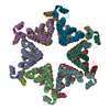
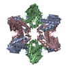
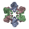
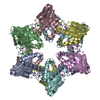
- EMDB-4906: Cryo-EM reconstruction of TssA protein from T6SS of Escherichia coli. -
+ Open data

- Basic information
| Entry | Database: EMDB / ID: EMD-4906 | |||||||||
|---|---|---|---|---|---|---|---|---|---|---|
| Title | Cryo-EM reconstruction of TssA protein from T6SS of Escherichia coli. | |||||||||
 Map data Map data | ||||||||||
 Sample Sample |
| |||||||||
 Keywords Keywords | T6SS / E. coli / TssA / cap / transport protein | |||||||||
| Function / homology | Type VI secretion system-associated, VCA0119 / Type VI secretion, EvfE, EvfF, ImpA, BimE, VC_A0119, VasJ / ImpA, N-terminal / ImpA, N-terminal, type VI secretion system / Putative type VI secretion protein Function and homology information Function and homology information | |||||||||
| Biological species |  | |||||||||
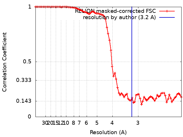
| Method | single particle reconstruction / cryo EM / Resolution: 3.2 Å | |||||||||
 Authors Authors | Nazarov S / Shneider M | |||||||||
| Funding support |  Switzerland, 1 items Switzerland, 1 items
| |||||||||
 Citation Citation | Journal: To Be Published Title: Cryo-EM structure of TssA protein from Type VI secretion system of E. coli. Authors: Nazarov S / Demurtas D / Shneider M / Basler M / Leiman P | |||||||||
| History |
|
- Structure visualization
| Movie |
Movie viewer |
|---|---|
| Structure viewer | EM map: SurfViewMolmilJmol/JSmol |
| Supplemental images |
- Downloads & links
-EMDB archive
| Map data | emd_4906.map.gz | 100.6 MB | EMDB map data format | |
|---|---|---|---|---|
| Header (meta data) | emd-4906-v30.xmlemd-4906.xml | 19.4 KB 19.4 KB | Display Display | EMDB header |
| FSC (resolution estimation) | emd_4906_fsc.xml | 7.8 KB | Display | FSC data file |
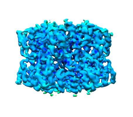
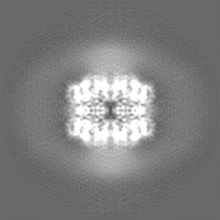
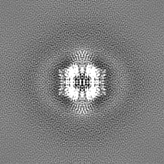
| Images |  emd_4906.png emd_4906.png | 144 KB | ||
| Masks | emd_4906_msk_1.map | 129.7 MB | Mask map | |
| Filedesc metadata | emd-4906.cif.gz | 6.2 KB | ||
| Others | emd_4906_additional_1.map.gzemd_4906_additional_2.map.gzemd_4906_half_map_1.map.gzemd_4906_half_map_2.map.gz | 37.8 MB 120.5 MB 95.6 MB 97.2 MB | ||
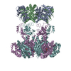
| Archive directory |  http://ftp.pdbj.org/pub/emdb/structures/EMD-4906ftp://ftp.pdbj.org/pub/emdb/structures/EMD-4906 http://ftp.pdbj.org/pub/emdb/structures/EMD-4906ftp://ftp.pdbj.org/pub/emdb/structures/EMD-4906 | HTTPS FTP |
-Related structure data
| Related structure data |  6rjuMC M: atomic model generated by this map C: citing same article ( |
|---|---|
| Similar structure data |
-Links
| EMDB pages | EMDB (EBI/PDBe) / EMDataResource |
|---|
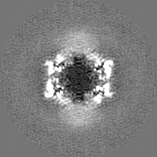
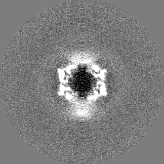
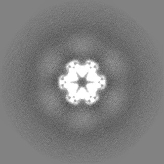
-Map
| File | Download / File: emd_4906.map.gz / Format: CCP4 / Size: 129.7 MB / Type: IMAGE STORED AS FLOATING POINT NUMBER (4 BYTES) | ||||||||||||||||||||||||||||||||||||||||||||||||||||||||||||
|---|---|---|---|---|---|---|---|---|---|---|---|---|---|---|---|---|---|---|---|---|---|---|---|---|---|---|---|---|---|---|---|---|---|---|---|---|---|---|---|---|---|---|---|---|---|---|---|---|---|---|---|---|---|---|---|---|---|---|---|---|---|
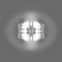
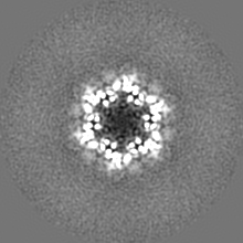
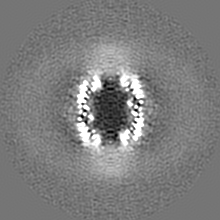
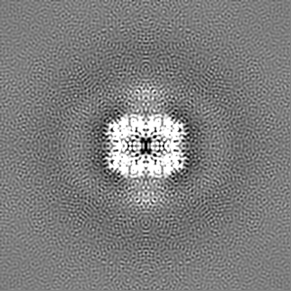
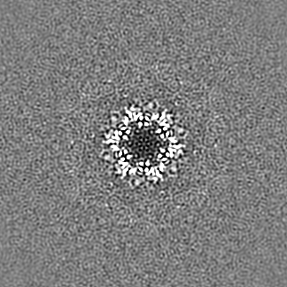
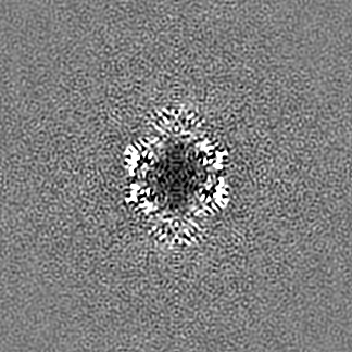
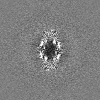
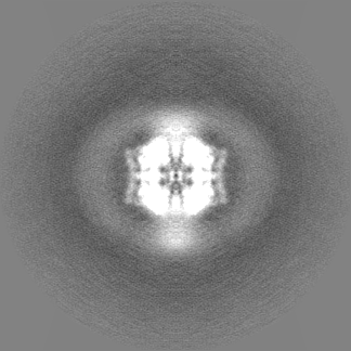
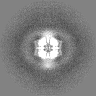
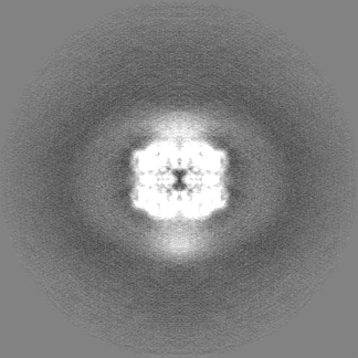
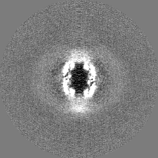
| Projections & slices | Image control
Images are generated by Spider. | ||||||||||||||||||||||||||||||||||||||||||||||||||||||||||||
| Voxel size | X=Y=Z: 1.058 Å | ||||||||||||||||||||||||||||||||||||||||||||||||||||||||||||
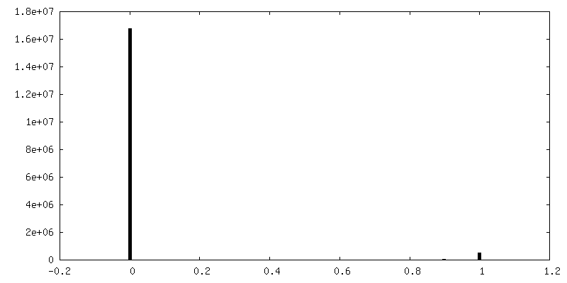
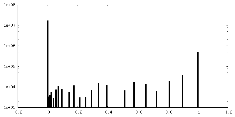
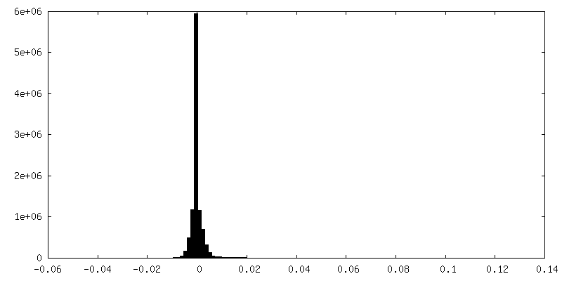
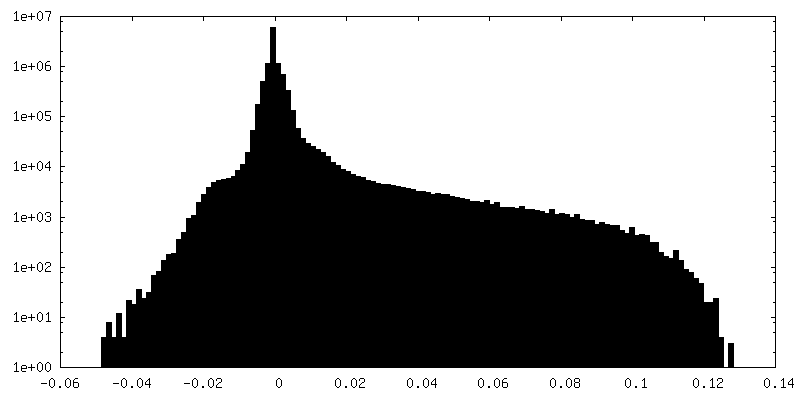
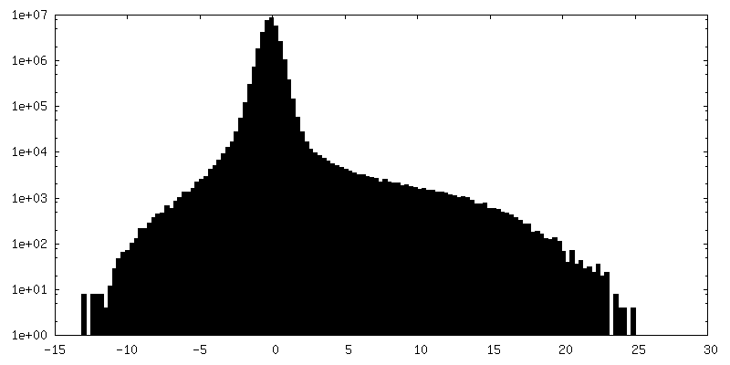
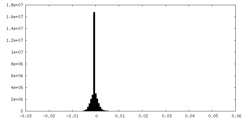
| Density |
| ||||||||||||||||||||||||||||||||||||||||||||||||||||||||||||
| Symmetry | Space group: 1 | ||||||||||||||||||||||||||||||||||||||||||||||||||||||||||||
| Details | EMDB XML:
CCP4 map header:
| ||||||||||||||||||||||||||||||||||||||||||||||||||||||||||||
 Z (Sec.)
 Y (Row.)
 X (Col.)


-Supplemental data
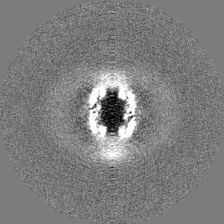
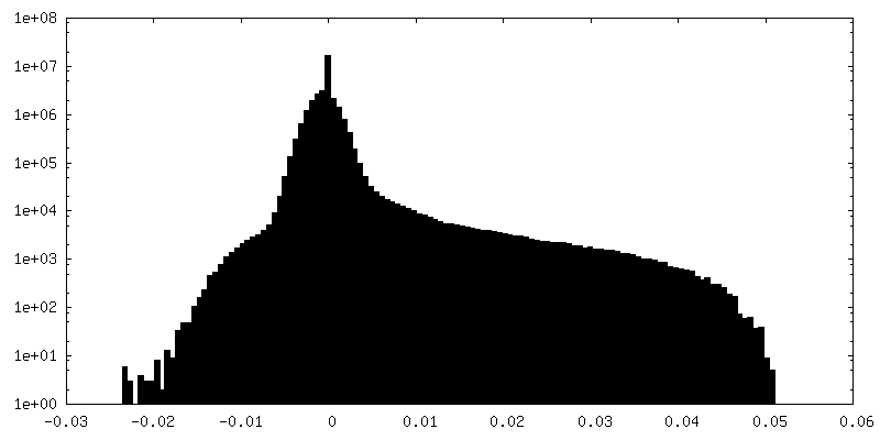
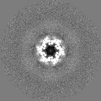
-Mask #1
| File | emd_4906_msk_1.map | ||||||||||||
|---|---|---|---|---|---|---|---|---|---|---|---|---|---|
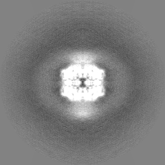
| Projections & Slices |
| ||||||||||||
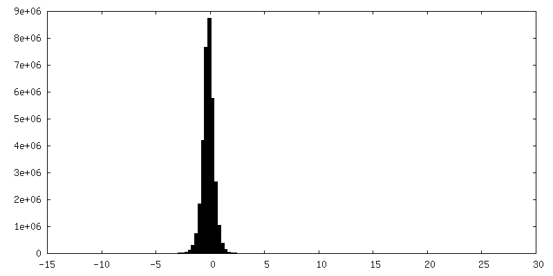
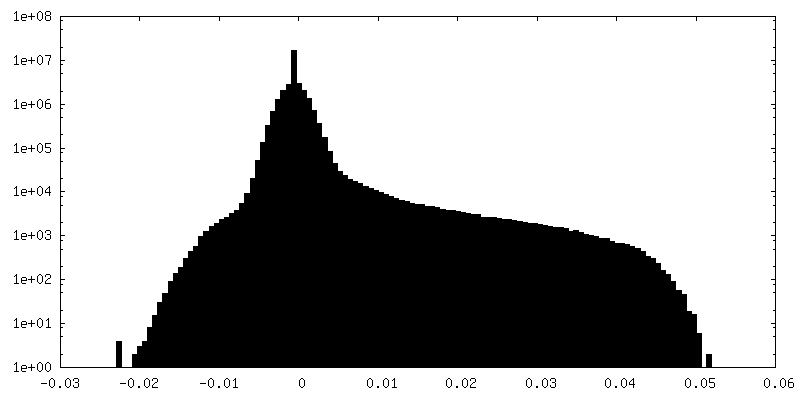
| Density Histograms |


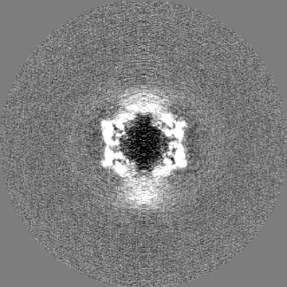
-Additional map: #1
| File | emd_4906_additional_1.map | ||||||||||||
|---|---|---|---|---|---|---|---|---|---|---|---|---|---|
| Projections & Slices |
| ||||||||||||
| Density Histograms |

-Additional map: #2
| File | emd_4906_additional_2.map | ||||||||||||
|---|---|---|---|---|---|---|---|---|---|---|---|---|---|
| Projections & Slices |
| ||||||||||||
| Density Histograms |

-Half map: #2
| File | emd_4906_half_map_1.map | ||||||||||||
|---|---|---|---|---|---|---|---|---|---|---|---|---|---|
| Projections & Slices |
| ||||||||||||
| Density Histograms |


-Half map: #1
| File | emd_4906_half_map_2.map | ||||||||||||
|---|---|---|---|---|---|---|---|---|---|---|---|---|---|
| Projections & Slices |
| ||||||||||||
| Density Histograms |
- Sample components
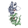
-Entire : C-terminal domain of T6SS protein TssA.
| Entire | Name: C-terminal domain of T6SS protein TssA. |
|---|---|
| Components |
|
-Supramolecule #1: C-terminal domain of T6SS protein TssA.
| Supramolecule | Name: C-terminal domain of T6SS protein TssA. / type: complex / ID: 1 / Parent: 0 / Macromolecule list: all |
|---|---|
| Source (natural) | Organism: |
| Molecular weight | Theoretical: 634 KDa |
-Macromolecule #1: Putative type VI secretion protein
| Macromolecule | Name: Putative type VI secretion protein / type: protein_or_peptide / ID: 1 / Number of copies: 1 / Enantiomer: LEVO |
|---|---|
| Source (natural) | Organism: |
| Molecular weight | Theoretical: 59.159789 KDa |
| Recombinant expression | Organism: |
| Sequence | String: MASIHSLLSA CQTTPRDVAE PAQVRIALWD KWLAPVTPDN PAGDDAGYDD DFQQMREEVN KLSGADAGIV SQLAEKLLTT RTKDIRVAT WYIWARLRQD GEKGLADGLE LLTGLLQRFG EHLHPQRSRA RKAALEWLCS ARILDSLSLY PEVVKADTLR I AGALWLAE ...String: MASIHSLLSA CQTTPRDVAE PAQVRIALWD KWLAPVTPDN PAGDDAGYDD DFQQMREEVN KLSGADAGIV SQLAEKLLTT RTKDIRVAT WYIWARLRQD GEKGLADGLE LLTGLLQRFG EHLHPQRSRA RKAALEWLCS ARILDSLSLY PEVVKADTLR I AGALWLAE QTFTDEASAP VLNGLYQALE NRLMKAGGVD AVVPQEAAAP APTVTSGSVM ALSAITSGQE LLSQARVLAK YL RDQPEGW LAAHRLMKSV RHDTLHQLPP LSADGRTRIA PPGPDRRASL KRLYLQQNWL SLLEQCDDMF ARGASHLWLD LQW YIHQAL LQTGKENYAA IIQYDLKGLL LRLPGLETLA FNDGMPFADD VTLSWIQQQV MECGERWAEE PSVTITAAPG DNDI LSLEP EALQIADNEG TEAALSWLQA RPGIQSDRSN WLLRLL(MSE)ARV AEQTGKNDLA LHLLAELDER ATRLTLSQWE P ELVFEVKA RRLKLLR(MSE)KS AKTESDRVRL QPD(MSE)EHLLAG LIAIDAARAA VLCNSGSS UniProtKB: Putative type VI secretion protein |
-Experimental details
-Structure determination
| Method | cryo EM |
|---|---|
 Processing Processing | single particle reconstruction |
| Aggregation state | particle |
-Sample preparation
| Buffer | pH: 7.5 |
|---|---|
| Vitrification | Cryogen name: ETHANE / Chamber humidity: 90 % / Chamber temperature: 293 K / Details: Leica EM GP2. |
- Electron microscopy
| Microscope | FEI TITAN KRIOS |
|---|---|
| Specialist optics | Energy filter - Name: GIF Quantum LS |
| Image recording | Film or detector model: GATAN K2 SUMMIT (4k x 4k) / Detector mode: COUNTING / Number real images: 3269 / Average electron dose: 60.0 e/Å2 |
| Electron beam | Acceleration voltage: 300 kV / Electron source:  FIELD EMISSION GUN FIELD EMISSION GUN |
| Electron optics | Illumination mode: FLOOD BEAM / Imaging mode: BRIGHT FIELD / Cs: 2.7 mm |
| Sample stage | Specimen holder model: FEI TITAN KRIOS AUTOGRID HOLDER / Cooling holder cryogen: NITROGEN |
| Experimental equipment |  Model: Titan Krios / Image courtesy: FEI Company |