National Institutes of Health/National Institute of General Medical Sciences (NIH/NIGMS)
R35 GM144282
米国
引用
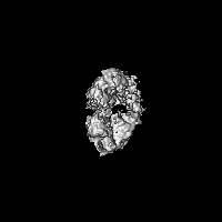
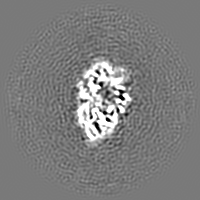
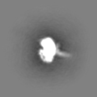
ジャーナル: Proc Natl Acad Sci U S A / 年: 2025 タイトル: Structural basis for dimerization and activation of UvrD-family helicases. 著者: Ankita Chadda / Binh Nguyen / Timothy M Lohman / Eric A Galburt / 要旨: UvrD-family helicases are superfamily 1A motor proteins that function during DNA replication, recombination, repair, and transcription. UvrD family monomers translocate along single-stranded (ss) DNA ...UvrD-family helicases are superfamily 1A motor proteins that function during DNA replication, recombination, repair, and transcription. UvrD family monomers translocate along single-stranded (ss) DNA but need to be activated by dimerization to unwind DNA in the absence of force or accessory factors. However, prior structural studies have only revealed monomeric complexes. Here, we report the first structures of a dimeric UvrD-family helicase, UvrD1, both free and bound to a DNA junction. In each structure, the dimer interface occurs between the 2B subdomains of each subunit. The apo UvrD1 dimer is observed in symmetric compact and extended forms indicating substantial flexibility. This symmetry is broken in the DNA-bound dimer complex with leading and trailing subunits adopting distinct conformations. Biochemical experiments reveal that the UvrD dimer shares the same 2B-2B interface. In contrast to the dimeric structures, an inactive, autoinhibited UvrD1 DNA-bound monomer structure reveals 2B subdomain-DNA contacts that are likely inhibitory. The major reorientation of the 2B subdomains that occurs upon UvrD1 dimerization prevents these duplex DNA interactions, thus relieving the autoinhibition. These structures reveal that the 2B subdomain serves a major regulatory role rather than participating directly in DNA unwinding.
 ムービー
ムービー コントローラー


 データを開く
 基本情報
基本情報
 マップデータ
 試料
試料 キーワード
キーワード 機能・相同性情報
 Mycobacterium tuberculosis (結核菌)
 データ登録者
 米国, 1件
  引用
引用 構造の表示
構造の表示
 ダウンロードとリンク
 emd_46797.png
 http://ftp.pdbj.org/pub/emdb/structures/EMD-46797


 Z (Sec.)
 Y (Row.)
 X (Col.)


 試料の構成要素
 解析
解析 電子顕微鏡法
 FIELD EMISSION GUN

