+ Open data
- Basic information
| Entry |  | |||||||||
|---|---|---|---|---|---|---|---|---|---|---|
| Title | Guillardia theta Fanzor (GtFz) State 1 | |||||||||
 Map data Map data | ||||||||||
 Sample Sample |
| |||||||||
 Keywords Keywords | Fanzor / Eukaryotic / RNA-guided / nuclease / Gene editing / RNA BINDING PROTEIN-RNA-DNA complex | |||||||||
| Function / homology |  Function and homology information Function and homology informationdetection of maltose stimulus / maltose transport complex / maltose binding / carbohydrate transport / maltose transport / maltodextrin transmembrane transport / carbohydrate transmembrane transporter activity / ATP-binding cassette (ABC) transporter complex, substrate-binding subunit-containing / ATP-binding cassette (ABC) transporter complex / cell chemotaxis ...detection of maltose stimulus / maltose transport complex / maltose binding / carbohydrate transport / maltose transport / maltodextrin transmembrane transport / carbohydrate transmembrane transporter activity / ATP-binding cassette (ABC) transporter complex, substrate-binding subunit-containing / ATP-binding cassette (ABC) transporter complex / cell chemotaxis / outer membrane-bounded periplasmic space / periplasmic space / DNA damage response / membrane Similarity search - Function | |||||||||
| Biological species |  | |||||||||
| Method | single particle reconstruction / cryo EM / Resolution: 4.7 Å | |||||||||
 Authors Authors | Xu P / Saito M / Zhang F | |||||||||
| Funding support |  United States, 1 items United States, 1 items
| |||||||||
 Citation Citation |  Journal: Cell / Year: 2024 Journal: Cell / Year: 2024Title: Structural insights into the diversity and DNA cleavage mechanism of Fanzor. Authors: Peiyu Xu / Makoto Saito / Guilhem Faure / Samantha Maguire / Samuel Chau-Duy-Tam Vo / Max E Wilkinson / Huihui Kuang / Bing Wang / William J Rice / Rhiannon K Macrae / Feng Zhang /  Abstract: Fanzor (Fz) is an ωRNA-guided endonuclease extensively found throughout the eukaryotic domain with unique gene editing potential. Here, we describe the structures of Fzs from three different ...Fanzor (Fz) is an ωRNA-guided endonuclease extensively found throughout the eukaryotic domain with unique gene editing potential. Here, we describe the structures of Fzs from three different organisms. We find that Fzs share a common ωRNA interaction interface, regardless of the length of the ωRNA, which varies considerably across species. The analysis also reveals Fz's mode of DNA recognition and unwinding capabilities as well as the presence of a non-canonical catalytic site. The structures demonstrate how protein conformations of Fz shift to allow the binding of double-stranded DNA to the active site within the R-loop. Mechanistically, examination of structures in different states shows that the conformation of the lid loop on the RuvC domain is controlled by the formation of the guide/DNA heteroduplex, regulating the activation of nuclease and DNA double-stranded displacement at the single cleavage site. Our findings clarify the mechanism of Fz, establishing a foundation for engineering efforts. | |||||||||
| History |
|
- Structure visualization
| Supplemental images |
|---|
- Downloads & links
-EMDB archive
| Map data |  emd_45516.map.gz emd_45516.map.gz | 167.9 MB |  EMDB map data format EMDB map data format | |
|---|---|---|---|---|
| Header (meta data) |  emd-45516-v30.xml emd-45516-v30.xml emd-45516.xml emd-45516.xml | 17.3 KB 17.3 KB | Display Display |  EMDB header EMDB header |
| Images |  emd_45516.png emd_45516.png | 30.6 KB | ||
| Filedesc metadata |  emd-45516.cif.gz emd-45516.cif.gz | 6.4 KB | ||
| Others |  emd_45516_additional_1.map.gz emd_45516_additional_1.map.gz emd_45516_half_map_1.map.gz emd_45516_half_map_1.map.gz emd_45516_half_map_2.map.gz emd_45516_half_map_2.map.gz | 87.1 MB 165.1 MB 165.1 MB | ||
| Archive directory |  http://ftp.pdbj.org/pub/emdb/structures/EMD-45516 http://ftp.pdbj.org/pub/emdb/structures/EMD-45516 ftp://ftp.pdbj.org/pub/emdb/structures/EMD-45516 ftp://ftp.pdbj.org/pub/emdb/structures/EMD-45516 | HTTPS FTP |
-Validation report
| Summary document |  emd_45516_validation.pdf.gz emd_45516_validation.pdf.gz | 1 MB | Display |  EMDB validaton report EMDB validaton report |
|---|---|---|---|---|
| Full document |  emd_45516_full_validation.pdf.gz emd_45516_full_validation.pdf.gz | 1 MB | Display | |
| Data in XML |  emd_45516_validation.xml.gz emd_45516_validation.xml.gz | 15.2 KB | Display | |
| Data in CIF |  emd_45516_validation.cif.gz emd_45516_validation.cif.gz | 18 KB | Display | |
| Arichive directory |  https://ftp.pdbj.org/pub/emdb/validation_reports/EMD-45516 https://ftp.pdbj.org/pub/emdb/validation_reports/EMD-45516 ftp://ftp.pdbj.org/pub/emdb/validation_reports/EMD-45516 ftp://ftp.pdbj.org/pub/emdb/validation_reports/EMD-45516 | HTTPS FTP |
-Related structure data
| Related structure data |  9cerMC  9cesC 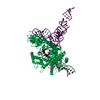 9cetC  9ceuC 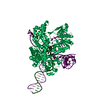 9cevC 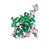 9cewC 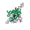 9cexC  9ceyC 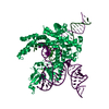 9cezC  9cf0C  9cf1C  9cf2C  9cf3C M: atomic model generated by this map C: citing same article ( |
|---|---|
| Similar structure data | Similarity search - Function & homology  F&H Search F&H Search |
- Links
Links
| EMDB pages |  EMDB (EBI/PDBe) / EMDB (EBI/PDBe) /  EMDataResource EMDataResource |
|---|---|
| Related items in Molecule of the Month |
- Map
Map
| File |  Download / File: emd_45516.map.gz / Format: CCP4 / Size: 178 MB / Type: IMAGE STORED AS FLOATING POINT NUMBER (4 BYTES) Download / File: emd_45516.map.gz / Format: CCP4 / Size: 178 MB / Type: IMAGE STORED AS FLOATING POINT NUMBER (4 BYTES) | ||||||||||||||||||||||||||||||||||||
|---|---|---|---|---|---|---|---|---|---|---|---|---|---|---|---|---|---|---|---|---|---|---|---|---|---|---|---|---|---|---|---|---|---|---|---|---|---|
| Projections & slices | Image control
Images are generated by Spider. | ||||||||||||||||||||||||||||||||||||
| Voxel size | X=Y=Z: 0.663 Å | ||||||||||||||||||||||||||||||||||||
| Density |
| ||||||||||||||||||||||||||||||||||||
| Symmetry | Space group: 1 | ||||||||||||||||||||||||||||||||||||
| Details | EMDB XML:
|
-Supplemental data
- Sample components
-Entire : GtFz-omegaRNA complex
| Entire | Name: GtFz-omegaRNA complex |
|---|---|
| Components |
|
-Supramolecule #1: GtFz-omegaRNA complex
| Supramolecule | Name: GtFz-omegaRNA complex / type: complex / ID: 1 / Parent: 0 / Macromolecule list: #1-#2 |
|---|---|
| Source (natural) | Organism:  |
-Macromolecule #1: Maltose/maltodextrin-binding periplasmic protein,Guillardia theta...
| Macromolecule | Name: Maltose/maltodextrin-binding periplasmic protein,Guillardia theta Fanzor1 type: protein_or_peptide / ID: 1 / Number of copies: 1 / Enantiomer: LEVO |
|---|---|
| Source (natural) | Organism:  |
| Molecular weight | Theoretical: 124.818938 KDa |
| Recombinant expression | Organism:  |
| Sequence | String: MKSSHHHHHH HHHHGSSMKI EEGKLVIWIN GDKGYNGLAE VGKKFEKDTG IKVTVEHPDK LEEKFPQVAA TGDGPDIIFW AHDRFGGYA QSGLLAEITP DKAFQDKLYP FTWDAVRYNG KLIAYPIAVE ALSLIYNKDL LPNPPKTWEE IPALDKELKA K GKSALMFN ...String: MKSSHHHHHH HHHHGSSMKI EEGKLVIWIN GDKGYNGLAE VGKKFEKDTG IKVTVEHPDK LEEKFPQVAA TGDGPDIIFW AHDRFGGYA QSGLLAEITP DKAFQDKLYP FTWDAVRYNG KLIAYPIAVE ALSLIYNKDL LPNPPKTWEE IPALDKELKA K GKSALMFN LQEPYFTWPL IAADGGYAFK YENGKYDIKD VGVDNAGAKA GLTFLVDLIK NKHMNADTDY SIAEAAFNKG ET AMTINGP WAWSNIDTSK VNYGVTVLPT FKGQPSKPFV GVLSAGINAA SPNKELAKEF LENYLLTDEG LEAVNKDKPL GAV ALKSYE EELAKDPRIA ATMENAQKGE IMPNIPQMSA FWYAVRTAVI NAASGRQTVD EALKDAQTNS SSNNNNNNNN NNLG IEENL YFQSNASNIR IVKRKAKGFF KCEDLVTIKD AVKAAHRIMS DASILVRSYY LRWFQSSYPL DSDDKELELE HFHIS MACS IVQGITRPPV RGVGPEQSVK IDVFNDMLDE YKRLYERAPN DKENETDLSL SHVLAYSIDN LLTAYKNNIE AHFSKY VKR FIRCDMLAKG FNKSEANRVA AIYTNAYIYD SSLDLEPDFM ERLGLEATSY SSLFPSKINK GGFPRVYDLK ANPWVYL PK MVMINQALET DFSSVEHKER RLLNPLPFYS SFVPMHIRID TSGLSQLLMT KDRLDDFKRS YLAEFGVSLN IKNKGDML A SFEKIFGRKA TSNREAGLYA TEMWSFLTNL KTCRQWKELD GVVRKNDPKG TQWMFDNAVV TDGVSISFQV IDNSMFGRK AFSGRKKRVA CQEANDEEDS KQVTREELKT SKLLGCDPGK RDILAITDGI KTICYTKGQR DMDTHKTIRL RTSLKRRRGC GLEEYETQV MNRFQKRSCH PEMFRRYACS RKRMEHMLLE CYSHPVFREF KFLVYNKTKS SEHRFMHRVL ETFKRPQTNL S KARCASGV MRMNALKEVQ RHGDIIIGWG NWGKNPNALR CSAGPTPGIG IRRRFESLFK TTTVPEHYTS QECPSCKGRC LR KATGNPI MRHHLLRCTN DSCCSRWWNR NVAGAFNILT RLLDGQTLSG NETTGDGLGG DDL UniProtKB: Maltose/maltodextrin-binding periplasmic protein, Cas12f1-like TNB domain-containing protein |
-Macromolecule #2: RNA (142-MER)
| Macromolecule | Name: RNA (142-MER) / type: rna / ID: 2 / Number of copies: 1 |
|---|---|
| Source (natural) | Organism:  |
| Molecular weight | Theoretical: 49.948816 KDa |
| Sequence | String: CACUAUCCGG UAACGAAACU ACCGGAGACG GGUUAGGAGG UGACGACCUC UAAAACCUAG AACUUAGAGU GCAAAAACGC CAUUACGAU UGUGAUGCCU AUUCAAGGGU GUCCCAAGUG UAAAAAGAAA GCACUCUAAG AGCAUUAAAC UCUACU |
-Macromolecule #3: ZINC ION
| Macromolecule | Name: ZINC ION / type: ligand / ID: 3 / Number of copies: 1 / Formula: ZN |
|---|---|
| Molecular weight | Theoretical: 65.409 Da |
-Experimental details
-Structure determination
| Method | cryo EM |
|---|---|
 Processing Processing | single particle reconstruction |
| Aggregation state | particle |
- Sample preparation
| Buffer | pH: 7.5 |
|---|---|
| Vitrification | Cryogen name: ETHANE-PROPANE |
- Electron microscopy
| Microscope | FEI TITAN KRIOS |
|---|---|
| Image recording | Film or detector model: GATAN K3 (6k x 4k) / Average electron dose: 51.41 e/Å2 |
| Electron beam | Acceleration voltage: 300 kV / Electron source:  FIELD EMISSION GUN FIELD EMISSION GUN |
| Electron optics | Illumination mode: FLOOD BEAM / Imaging mode: BRIGHT FIELD / Cs: 2.7 mm / Nominal defocus max: 2.6 µm / Nominal defocus min: 0.5 µm |
| Experimental equipment |  Model: Titan Krios / Image courtesy: FEI Company |
- Image processing
| Startup model | Type of model: NONE |
|---|---|
| Final reconstruction | Resolution.type: BY AUTHOR / Resolution: 4.7 Å / Resolution method: FSC 0.143 CUT-OFF / Number images used: 10507 |
| Initial angle assignment | Type: RANDOM ASSIGNMENT |
| Final angle assignment | Type: MAXIMUM LIKELIHOOD |
 Movie
Movie Controller


 Z (Sec.)
 Y (Row.)
 X (Col.)


