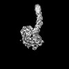
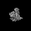
 Movie
Movie Controller


+ Open data

- Basic information
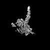
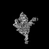
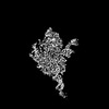
| Entry | Database: PDB / ID: 9cf0 | |||||||||
|---|---|---|---|---|---|---|---|---|---|---|
| Title | Parasitella parasitica Fanzor (PpFz) State 1 | |||||||||
 Components Components |
| |||||||||
 Keywords Keywords | RNA BINDING PROTEIN/ISOMERASE/DNA / Fanzor / Eukaryotic / RNA-guided / nuclease / Gene editing / RNA BINDING PROTEIN-ISOMERASE-DNA complex | |||||||||
| Function / homology |  Function and homology information Function and homology informationSet3 complex / negative regulation of meiotic nuclear division / ascospore formation / positive regulation of meiotic nuclear division / cyclosporin A binding / detection of maltose stimulus / maltose transport complex / carbohydrate transport / carbohydrate transmembrane transporter activity / maltose binding ...Set3 complex / negative regulation of meiotic nuclear division / ascospore formation / positive regulation of meiotic nuclear division / cyclosporin A binding / detection of maltose stimulus / maltose transport complex / carbohydrate transport / carbohydrate transmembrane transporter activity / maltose binding / maltose transport / maltodextrin transmembrane transport / ATP-binding cassette (ABC) transporter complex, substrate-binding subunit-containing / ATP-binding cassette (ABC) transporter complex / cellular response to starvation / peptidylprolyl isomerase / peptidyl-prolyl cis-trans isomerase activity / cell chemotaxis / mitochondrial intermembrane space / outer membrane-bounded periplasmic space / protein transport / protein folding / periplasmic space / mRNA binding / DNA damage response / mitochondrion / membrane / nucleus / cytoplasm Similarity search - Function | |||||||||
| Biological species |  synthetic construct (others)  Parasitella parasitica (fungus) Parasitella parasitica (fungus) | |||||||||
| Method | ELECTRON MICROSCOPY / single particle reconstruction / cryo EM / Resolution: 3.47 Å | |||||||||
 Authors Authors | Xu, P. / Saito, M. / Zhang, F. | |||||||||
| Funding support |  United States, 1items United States, 1items
| |||||||||
 Citation Citation | Journal: Cell / Year: 2024 Title: Structural insights into the diversity and DNA cleavage mechanism of Fanzor. Authors: Peiyu Xu / Makoto Saito / Guilhem Faure / Samantha Maguire / Samuel Chau-Duy-Tam Vo / Max E Wilkinson / Huihui Kuang / Bing Wang / William J Rice / Rhiannon K Macrae / Feng Zhang / Abstract: Fanzor (Fz) is an ωRNA-guided endonuclease extensively found throughout the eukaryotic domain with unique gene editing potential. Here, we describe the structures of Fzs from three different ...Fanzor (Fz) is an ωRNA-guided endonuclease extensively found throughout the eukaryotic domain with unique gene editing potential. Here, we describe the structures of Fzs from three different organisms. We find that Fzs share a common ωRNA interaction interface, regardless of the length of the ωRNA, which varies considerably across species. The analysis also reveals Fz's mode of DNA recognition and unwinding capabilities as well as the presence of a non-canonical catalytic site. The structures demonstrate how protein conformations of Fz shift to allow the binding of double-stranded DNA to the active site within the R-loop. Mechanistically, examination of structures in different states shows that the conformation of the lid loop on the RuvC domain is controlled by the formation of the guide/DNA heteroduplex, regulating the activation of nuclease and DNA double-stranded displacement at the single cleavage site. Our findings clarify the mechanism of Fz, establishing a foundation for engineering efforts. | |||||||||
| History |
|
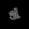
- Structure visualization
| Structure viewer | Molecule: MolmilJmol/JSmol |
|---|
- Downloads & links
-Download
| PDBx/mmCIF format | 9cf0.cif.gz | 453.9 KB | Display | PDBx/mmCIF format |
|---|---|---|---|---|
| PDB format | pdb9cf0.ent.gz | 296.3 KB | Display | PDB format |
| PDBx/mmJSON format | 9cf0.json.gz | Tree view | PDBx/mmJSON format | |
| Others |  Other downloads Other downloads |
-Validation report
| Arichive directory | https://data.pdbj.org/pub/pdb/validation_reports/cf/9cf0ftp://data.pdbj.org/pub/pdb/validation_reports/cf/9cf0 | HTTPS FTP |
|---|
-Related structure data
| Related structure data | 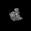 45525MC  9cerC  9cesC 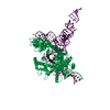 9cetC  9ceuC 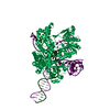 9cevC 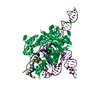 9cewC 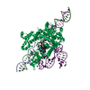 9cexC  9ceyC 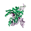 9cezC  9cf1C 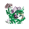 9cf2C 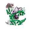 9cf3C M: map data used to model this data C: citing same article ( |
|---|---|
| Similar structure data |

-Links
 PDBj
PDBj


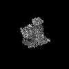
- Assembly
| Deposited unit | 
|
|---|---|
| 1 |
|
-Components
-Protein , 2 types, 2 molecules CP
| #1: Protein | Mass: 17411.670 Da / Num. of mol.: 1 Source method: isolated from a genetically manipulated source Source: (gene. exp.) Gene: CPR1, CPH1, CYP1, SCC1, YDR155C, YD8358.10C / Production host: |
|---|---|
| #3: Protein | Mass: 144413.969 Da / Num. of mol.: 1 Source method: isolated from a genetically manipulated source Source: (gene. exp.) Parasitella parasitica (fungus) / Gene: malE, b4034, JW3994, PARPA_09356.1 scaffold 36248 / Production host: |
-DNA chain , 2 types, 2 molecules NT
| #2: DNA chain | Mass: 17449.246 Da / Num. of mol.: 1 Source method: isolated from a genetically manipulated source Source: (gene. exp.) synthetic construct (others) / Production host: synthetic construct (others) |
|---|---|
| #4: DNA chain | Mass: 17675.332 Da / Num. of mol.: 1 Source method: isolated from a genetically manipulated source Source: (gene. exp.) synthetic construct (others) / Production host: synthetic construct (others) |
-RNA chain / Non-polymers , 2 types, 2 molecules W
| #5: RNA chain | Mass: 19514.656 Da / Num. of mol.: 1 Source method: isolated from a genetically manipulated source Source: (gene. exp.) Parasitella parasitica (fungus) / Production host: |
|---|---|
| #6: Chemical | ChemComp-ZN /  Mass: 65.409 Da / Num. of mol.: 1 / Source method: obtained synthetically / Formula: Zn Mass: 65.409 Da / Num. of mol.: 1 / Source method: obtained synthetically / Formula: Zn |
-Details
| Has ligand of interest | N |
|---|
-Experimental details
-Experiment
| Experiment | Method: ELECTRON MICROSCOPY |
|---|---|
| EM experiment | Aggregation state: PARTICLE / 3D reconstruction method: single particle reconstruction |
- Sample preparation
| Component | Name: Ternary complex of PpFz1-omegaRNA-DNA / Type: COMPLEX / Entity ID: #1-#5 / Source: RECOMBINANT |
|---|---|
| Source (natural) | Organism: Parasitella parasitica (fungus) |
| Source (recombinant) | Organism: |
| Buffer solution | pH: 7.4 |
| Specimen | Embedding applied: NO / Shadowing applied: NO / Staining applied: NO / Vitrification applied: YES |
| Vitrification | Cryogen name: ETHANE-PROPANE |
- Electron microscopy imaging
| Experimental equipment |  Model: Titan Krios / Image courtesy: FEI Company |
|---|---|
| Microscopy | Model: FEI TITAN KRIOS |
| Electron gun | Electron source:  FIELD EMISSION GUN / Accelerating voltage: 300 kV / Illumination mode: OTHER FIELD EMISSION GUN / Accelerating voltage: 300 kV / Illumination mode: OTHER |
| Electron lens | Mode: OTHER / Nominal defocus max: 2500 nm / Nominal defocus min: 500 nm |
| Image recording | Electron dose: 48.47 e/Å2 / Film or detector model: GATAN K3 (6k x 4k) |
- Processing
| EM software | Name: PHENIX / Version: 1.21_5207 / Category: model refinement | ||||||||||||||||||||||||
|---|---|---|---|---|---|---|---|---|---|---|---|---|---|---|---|---|---|---|---|---|---|---|---|---|---|
| CTF correction | Type: NONE | ||||||||||||||||||||||||
| 3D reconstruction | Resolution: 3.47 Å / Resolution method: FSC 0.143 CUT-OFF / Num. of particles: 15797 / Symmetry type: POINT | ||||||||||||||||||||||||
| Refinement | Cross valid method: NONE Stereochemistry target values: GeoStd + Monomer Library + CDL v1.2 | ||||||||||||||||||||||||
| Displacement parameters | Biso mean: 98.01 Å2 | ||||||||||||||||||||||||
| Refine LS restraints |
|
