- EMDB-45430: Human Mitochondrial LONP1 Degrading Casein, ATP-bound closed form -
+
Open data
ID or keywords:
Loading...
-
Basic information
Entry
Database: EMDB / ID: EMD-45430
Title
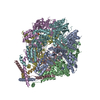
Human Mitochondrial LONP1 Degrading Casein, ATP-bound closed form
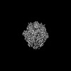
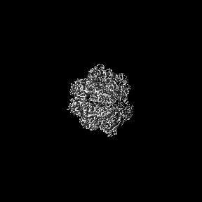
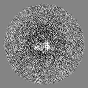
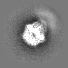
Map data
Sharpened map of LONP1 enzymatic assembly
Sample
Complex: LONP1 degrading casein
Protein or peptide: Lon protease homolog, mitochondrial
Protein or peptide: Bound substrate segment undergoing translocation and subsequent degradation
Ligand: ADENOSINE-5'-TRIPHOSPHATE
Ligand: MAGNESIUM ION
Ligand: ADENOSINE-5'-DIPHOSPHATE
Keywords
ATPase / protease / HYDROLASE
Function / homology
Function and homology information
oxidation-dependent protein catabolic process / response to aluminum ion / PH domain binding / mitochondrial protein catabolic process / endopeptidase La / G-quadruplex DNA binding / mitochondrial DNA metabolic process / : / ATP-dependent peptidase activity / protein quality control for misfolded or incompletely synthesized proteins ...oxidation-dependent protein catabolic process / response to aluminum ion / PH domain binding / mitochondrial protein catabolic process / endopeptidase La / G-quadruplex DNA binding / mitochondrial DNA metabolic process / : / ATP-dependent peptidase activity / protein quality control for misfolded or incompletely synthesized proteins / mitochondrial nucleoid / insulin receptor substrate binding / Mitochondrial unfolded protein response (UPRmt) / chaperone-mediated protein complex assembly / response to hormone / DNA polymerase binding / Mitochondrial protein degradation / negative regulation of insulin receptor signaling pathway / proteolysis involved in protein catabolic process / mitochondrion organization / protein catabolic process / ADP binding / single-stranded DNA binding / cellular response to oxidative stress / sequence-specific DNA binding / response to hypoxia / single-stranded RNA binding / mitochondrial matrix / serine-type endopeptidase activity / ATP hydrolysis activity / mitochondrion / nucleoplasm / ATP binding / identical protein binding / membrane / cytosol Similarity search - Function
Lon protease homologue, chloroplastic/mitochondrial / : / Lon protease, bacterial/eukaryotic-type / Lon protease AAA+ ATPase lid domain / Peptidase S16, active site / ATP-dependent serine proteases, lon family, serine active site. / Lon proteolytic domain profile. / Peptidase S16, Lon proteolytic domain / Lon protease / Lon protease (S16) C-terminal proteolytic domain ...Lon protease homologue, chloroplastic/mitochondrial / : / Lon protease, bacterial/eukaryotic-type / Lon protease AAA+ ATPase lid domain / Peptidase S16, active site / ATP-dependent serine proteases, lon family, serine active site. / Lon proteolytic domain profile. / Peptidase S16, Lon proteolytic domain / Lon protease / Lon protease (S16) C-terminal proteolytic domain / Lon N-terminal domain profile. / Lon protease, N-terminal domain / Lon protease, N-terminal domain superfamily / ATP-dependent protease La (LON) substrate-binding domain / Found in ATP-dependent protease La (LON) / PUA-like superfamily / ATPase family associated with various cellular activities (AAA) / ATPase, AAA-type, core / Ribosomal protein S5 domain 2-type fold, subgroup / Ribosomal protein S5 domain 2-type fold / ATPases associated with a variety of cellular activities / AAA+ ATPase domain / P-loop containing nucleoside triphosphate hydrolase Similarity search - Domain/homology
National Institutes of Health/National Institute of General Medical Sciences (NIH/NIGMS)
F32GM145143
United States
National Institutes of Health/National Institute of Neurological Disorders and Stroke (NIH/NINDS)
NS095892
United States
Citation
Journal: bioRxiv / Year: 2024 Title: Structural and mechanistic studies on human LONP1 redefine the hand-over-hand translocation mechanism. Authors: Jeffrey T Mindrebo / Gabriel C Lander / Abstract: AAA+ enzymes use energy from ATP hydrolysis to remodel diverse cellular targets. Structures of substrate-bound AAA+ complexes suggest that these enzymes employ a conserved hand-over-hand mechanism to ...AAA+ enzymes use energy from ATP hydrolysis to remodel diverse cellular targets. Structures of substrate-bound AAA+ complexes suggest that these enzymes employ a conserved hand-over-hand mechanism to thread substrates through their central pore. However, the fundamental aspects of the mechanisms governing motor function and substrate processing within specific AAA+ families remain unresolved. We used cryo-electron microscopy to structurally interrogate reaction intermediates from in vitro biochemical assays to inform the underlying regulatory mechanisms of the human mitochondrial AAA+ protease, LONP1. Our results demonstrate that substrate binding allosterically regulates proteolytic activity, and that LONP1 can adopt a configuration conducive to substrate translocation even when the ATPases are bound to ADP. These results challenge the conventional understanding of the hand-over-hand translocation mechanism, giving rise to an alternative model that aligns more closely with biochemical and biophysical data on related enzymes like ClpX, ClpA, the 26S proteasome, and Lon protease.
In the structure databanks used in Yorodumi, some data are registered as the other names, "COVID-19 virus" and "2019-nCoV". Here are the details of the virus and the list of structure data.
Jan 31, 2019. EMDB accession codes are about to change! (news from PDBe EMDB page)
EMDB accession codes are about to change! (news from PDBe EMDB page)
The allocation of 4 digits for EMDB accession codes will soon come to an end. Whilst these codes will remain in use, new EMDB accession codes will include an additional digit and will expand incrementally as the available range of codes is exhausted. The current 4-digit format prefixed with “EMD-” (i.e. EMD-XXXX) will advance to a 5-digit format (i.e. EMD-XXXXX), and so on. It is currently estimated that the 4-digit codes will be depleted around Spring 2019, at which point the 5-digit format will come into force.
The EM Navigator/Yorodumi systems omit the EMD- prefix.
Related info.:Q: What is EMD? / ID/Accession-code notation in Yorodumi/EM Navigator
Yorodumi is a browser for structure data from EMDB, PDB, SASBDB, etc.
This page is also the successor to EM Navigator detail page, and also detail information page/front-end page for Omokage search.
The word "yorodu" (or yorozu) is an old Japanese word meaning "ten thousand". "mi" (miru) is to see.
Related info.:EMDB / PDB / SASBDB / Comparison of 3 databanks / Yorodumi Search / Aug 31, 2016. New EM Navigator & Yorodumi / Yorodumi Papers / Jmol/JSmol / Function and homology information / Changes in new EM Navigator and Yorodumi
 Movie
Movie Controller

 Yorodumi
 Open data
 Basic information
 Map data
 Sample
 Keywords
 Function and homology information
 Homo sapiens (human) /
 Authors
 United States, 2 items
  Citation
 Structure visualization
 Downloads & links
 emd_45430.png
 http://ftp.pdbj.org/pub/emdb/structures/EMD-45430
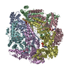


 Z (Sec.)
 Y (Row.)
 X (Col.)


 Sample components


 Processing
 Electron microscopy
 FIELD EMISSION GUN

