Movie
Movie Controller


+ Open data

- Basic information
| Entry | 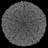 | |||||||||
|---|---|---|---|---|---|---|---|---|---|---|
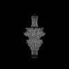
| Title | Pseudomonas phage DEV 5-fold vertex (major coat protein) | |||||||||
 Map data Map data | ||||||||||
 Sample Sample |
| |||||||||
 Keywords Keywords | virion coat / complex / STRUCTURAL PROTEIN / gp77 | |||||||||
| Function / homology | Major coat protein Function and homology information Function and homology information | |||||||||
| Biological species |  Pseudomonas phage vB_PaeP_DEV (virus) Pseudomonas phage vB_PaeP_DEV (virus) | |||||||||
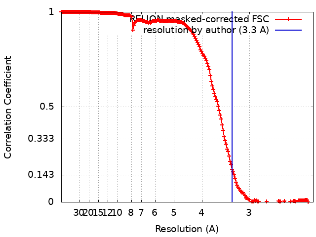
| Method | single particle reconstruction / cryo EM / Resolution: 3.3 Å | |||||||||
 Authors Authors | Iglesias SM / Hou CFD / Li F / Cingolani G | |||||||||
| Funding support |  United States, 2 items United States, 2 items
| |||||||||
 Citation Citation | Journal: To Be Published Title: Integrative structural analysis of Pseudomonas phage DEV reveals a genome ejection motor Authors: Lokareddy R / Hou CFD / Forti F / Iglesias SM / Li F / Pavlenok M / Niederweis M / Briani F / Cingolani G | |||||||||
| History |
|
- Structure visualization
| Supplemental images |
|---|

- Downloads & links
-EMDB archive
| Map data | emd_44518.map.gz | 472.9 MB | EMDB map data format | |
|---|---|---|---|---|
| Header (meta data) | emd-44518-v30.xmlemd-44518.xml | 13.6 KB 13.6 KB | Display Display | EMDB header |
| FSC (resolution estimation) | emd_44518_fsc.xml | 18.1 KB | Display | FSC data file |
| Images |  emd_44518.png emd_44518.png | 104.1 KB | ||
| Masks | emd_44518_msk_1.map | 512 MB | Mask map | |
| Filedesc metadata | emd-44518.cif.gz | 5.4 KB | ||
| Others | emd_44518_half_map_1.map.gzemd_44518_half_map_2.map.gz | 412.9 MB 411.4 MB | ||
| Archive directory |  http://ftp.pdbj.org/pub/emdb/structures/EMD-44518ftp://ftp.pdbj.org/pub/emdb/structures/EMD-44518 http://ftp.pdbj.org/pub/emdb/structures/EMD-44518ftp://ftp.pdbj.org/pub/emdb/structures/EMD-44518 | HTTPS FTP |
-Validation report
| Summary document | emd_44518_validation.pdf.gz | 1.3 MB | Display | EMDB validaton report |
|---|---|---|---|---|
| Full document | emd_44518_full_validation.pdf.gz | 1.3 MB | Display | |
| Data in XML | emd_44518_validation.xml.gz | 26.3 KB | Display | |
| Data in CIF | emd_44518_validation.cif.gz | 35.4 KB | Display | |
| Arichive directory | https://ftp.pdbj.org/pub/emdb/validation_reports/EMD-44518ftp://ftp.pdbj.org/pub/emdb/validation_reports/EMD-44518 | HTTPS FTP |
-Related structure data
| Related structure data | 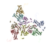 9bgnMC 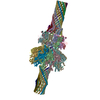 9bgmC M: atomic model generated by this map C: citing same article ( |
|---|---|
| Similar structure data |
-Links
| EMDB pages | EMDB (EBI/PDBe) / EMDataResource |
|---|
-Map
| File | Download / File: emd_44518.map.gz / Format: CCP4 / Size: 512 MB / Type: IMAGE STORED AS FLOATING POINT NUMBER (4 BYTES) | ||||||||||||||||||||||||||||||||||||
|---|---|---|---|---|---|---|---|---|---|---|---|---|---|---|---|---|---|---|---|---|---|---|---|---|---|---|---|---|---|---|---|---|---|---|---|---|---|
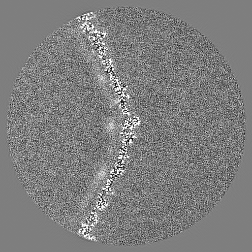
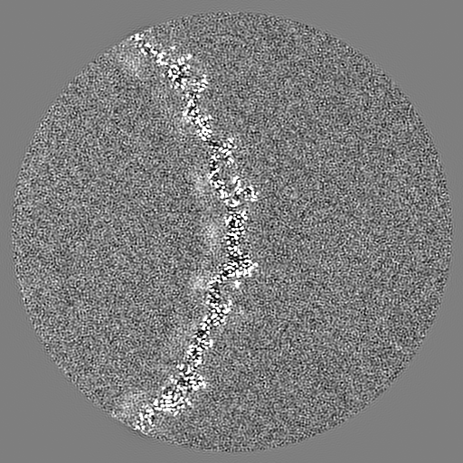
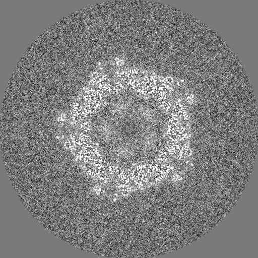
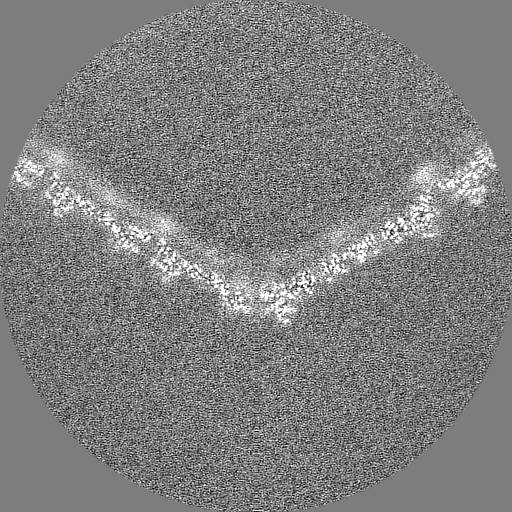
| Projections & slices | Image control
Images are generated by Spider. | ||||||||||||||||||||||||||||||||||||
| Voxel size | X=Y=Z: 1.12 Å | ||||||||||||||||||||||||||||||||||||
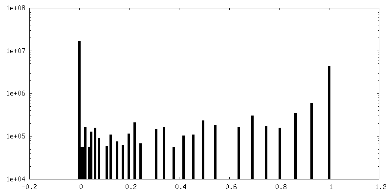
| Density |
| ||||||||||||||||||||||||||||||||||||
| Symmetry | Space group: 1 | ||||||||||||||||||||||||||||||||||||
| Details | EMDB XML:
|
 X (Sec.)
 Y (Row.)
 Z (Col.)


-Supplemental data
-Mask #1
| File | emd_44518_msk_1.map | ||||||||||||
|---|---|---|---|---|---|---|---|---|---|---|---|---|---|
| Projections & Slices |
| ||||||||||||
| Density Histograms |


-Half map: #1
| File | emd_44518_half_map_1.map | ||||||||||||
|---|---|---|---|---|---|---|---|---|---|---|---|---|---|
| Projections & Slices |
| ||||||||||||
| Density Histograms |


-Half map: #2
| File | emd_44518_half_map_2.map | ||||||||||||
|---|---|---|---|---|---|---|---|---|---|---|---|---|---|
| Projections & Slices |
| ||||||||||||
| Density Histograms |


- Sample components
-Entire : Major coat protein of Pseudomonas phage DEV
| Entire | Name: Major coat protein of Pseudomonas phage DEV |
|---|---|
| Components |
|
-Supramolecule #1: Major coat protein of Pseudomonas phage DEV
| Supramolecule | Name: Major coat protein of Pseudomonas phage DEV / type: complex / ID: 1 / Parent: 0 / Macromolecule list: all |
|---|---|
| Source (natural) | Organism: Pseudomonas phage vB_PaeP_DEV (virus) |
-Macromolecule #1: gp77 major coat protein
| Macromolecule | Name: gp77 major coat protein / type: protein_or_peptide / ID: 1 / Number of copies: 9 / Enantiomer: LEVO |
|---|---|
| Source (natural) | Organism: Pseudomonas phage vB_PaeP_DEV (virus) |
| Molecular weight | Theoretical: 44.114113 KDa |
| Recombinant expression | Organism:  Pseudomonas (RNA similarity group I) Pseudomonas (RNA similarity group I) |
| Sequence | String: MAGPVDNIKP MKYNDPANGV ESSIGPQIHT RYWYKRALID AAKEAYFGQL ADTFSMPKHY GKEIVRLHYI PLLDDRNVND QGIDASGAT IANGNLYGSS RDVGNITAKM PTLTEIGGRV NRVGFKRVEI KGKLEKYGFF REYTQEQLDF DSDPAMEGHV T TEMVKGAN ...String: MAGPVDNIKP MKYNDPANGV ESSIGPQIHT RYWYKRALID AAKEAYFGQL ADTFSMPKHY GKEIVRLHYI PLLDDRNVND QGIDASGAT IANGNLYGSS RDVGNITAKM PTLTEIGGRV NRVGFKRVEI KGKLEKYGFF REYTQEQLDF DSDPAMEGHV T TEMVKGAN EITEDLLQID LLNSAGTVRY PGAATSDAEV DASTEVTYDS LMRLRLDLDN ARAPTKIKMI TGTRMIDTRT VG NARALYV GSDLVPTIEA MKDNHGNPAF IPIEKYAAGG ATMHGEVGQL GRFRVIVNPQ MMHWAGVGKA VDPNDQVPMH ESG GKYSVF PMLCVASEAF TTVGFATDGK NVKFKIITKR PGEATADRSD PYGEMGFMSI KWYYGFMVFR PEWIALLKTV ARL UniProtKB: Major coat protein |
-Experimental details
-Structure determination
| Method | cryo EM |
|---|---|
 Processing Processing | single particle reconstruction |
| Aggregation state | particle |
-Sample preparation
| Buffer | pH: 7.5 |
|---|---|
| Vitrification | Cryogen name: ETHANE |
- Electron microscopy
| Microscope | FEI TITAN KRIOS |
|---|---|
| Image recording | Film or detector model: GATAN K3 (6k x 4k) / Average electron dose: 50.0 e/Å2 |
| Electron beam | Acceleration voltage: 300 kV / Electron source:  FIELD EMISSION GUN FIELD EMISSION GUN |
| Electron optics | Illumination mode: FLOOD BEAM / Imaging mode: BRIGHT FIELD / Nominal defocus max: 1.6 µm / Nominal defocus min: 0.8 µm |
| Experimental equipment |  Model: Titan Krios / Image courtesy: FEI Company |