+ Open data
- Basic information
| Entry |  | |||||||||
|---|---|---|---|---|---|---|---|---|---|---|
| Title | Structure of wild type human PSS1 | |||||||||
 Map data Map data | ||||||||||
 Sample Sample |
| |||||||||
 Keywords Keywords | MEMBRANE PROTEIN | |||||||||
| Function / homology |  Function and homology information Function and homology informationL-serine-phosphatidylethanolamine phosphatidyltransferase / L-serine-phosphatidylethanolamine phosphatidyltransferase activity / L-serine-phosphatidylcholine phosphatidyltransferase activity / Synthesis of PS / phosphatidylserine biosynthetic process / transferase activity / endoplasmic reticulum membrane / membrane Similarity search - Function | |||||||||
| Biological species |  Homo sapiens (human) Homo sapiens (human) | |||||||||
| Method | single particle reconstruction / cryo EM / Resolution: 2.7 Å | |||||||||
 Authors Authors | Long T / Li X | |||||||||
| Funding support |  United States, 1 items United States, 1 items
| |||||||||
 Citation Citation |  Journal: Cell / Year: 2024 Journal: Cell / Year: 2024Title: Molecular insights into human phosphatidylserine synthase 1 reveal its inhibition promotes LDL uptake. Authors: Tao Long / Dongyu Li / Goncalo Vale / Yaoyukun Jiang / Philip Schmiege / Zhongyue J Yang / Jeffrey G McDonald / Xiaochun Li /  Abstract: In mammalian cells, two phosphatidylserine (PS) synthases drive PS synthesis. Gain-of-function mutations in the Ptdss1 gene lead to heightened PS production, causing Lenz-Majewski syndrome (LMS). ...In mammalian cells, two phosphatidylserine (PS) synthases drive PS synthesis. Gain-of-function mutations in the Ptdss1 gene lead to heightened PS production, causing Lenz-Majewski syndrome (LMS). Recently, pharmacological inhibition of PSS1 has been shown to suppress tumorigenesis. Here, we report the cryo-EM structures of wild-type human PSS1 (PSS1), the LMS-causing Pro269Ser mutant (PSS1), and PSS1 in complex with its inhibitor DS55980254. PSS1 contains 10 transmembrane helices (TMs), with TMs 4-8 forming a catalytic core in the luminal leaflet. These structures revealed a working mechanism of PSS1 akin to the postulated mechanisms of the membrane-bound O-acyltransferase family. Additionally, we showed that both PS and DS55980254 can allosterically inhibit PSS1 and that inhibition by DS55980254 activates the SREBP pathways, thus enhancing the expression of LDL receptors and increasing cellular LDL uptake. This work uncovers a mechanism of mammalian PS synthesis and suggests that selective PSS1 inhibitors have the potential to lower blood cholesterol levels. | |||||||||
| History |
|
- Structure visualization
| Supplemental images |
|---|
- Downloads & links
-EMDB archive
| Map data |  emd_44178.map.gz emd_44178.map.gz | 97 MB |  EMDB map data format EMDB map data format | |
|---|---|---|---|---|
| Header (meta data) |  emd-44178-v30.xml emd-44178-v30.xml emd-44178.xml emd-44178.xml | 15.3 KB 15.3 KB | Display Display |  EMDB header EMDB header |
| Images |  emd_44178.png emd_44178.png | 36.9 KB | ||
| Filedesc metadata |  emd-44178.cif.gz emd-44178.cif.gz | 5.9 KB | ||
| Others |  emd_44178_half_map_1.map.gz emd_44178_half_map_1.map.gz emd_44178_half_map_2.map.gz emd_44178_half_map_2.map.gz | 95.3 MB 95.3 MB | ||
| Archive directory |  http://ftp.pdbj.org/pub/emdb/structures/EMD-44178 http://ftp.pdbj.org/pub/emdb/structures/EMD-44178 ftp://ftp.pdbj.org/pub/emdb/structures/EMD-44178 ftp://ftp.pdbj.org/pub/emdb/structures/EMD-44178 | HTTPS FTP |
-Validation report
| Summary document |  emd_44178_validation.pdf.gz emd_44178_validation.pdf.gz | 855.8 KB | Display |  EMDB validaton report EMDB validaton report |
|---|---|---|---|---|
| Full document |  emd_44178_full_validation.pdf.gz emd_44178_full_validation.pdf.gz | 855.4 KB | Display | |
| Data in XML |  emd_44178_validation.xml.gz emd_44178_validation.xml.gz | 13.2 KB | Display | |
| Data in CIF |  emd_44178_validation.cif.gz emd_44178_validation.cif.gz | 15.2 KB | Display | |
| Arichive directory |  https://ftp.pdbj.org/pub/emdb/validation_reports/EMD-44178 https://ftp.pdbj.org/pub/emdb/validation_reports/EMD-44178 ftp://ftp.pdbj.org/pub/emdb/validation_reports/EMD-44178 ftp://ftp.pdbj.org/pub/emdb/validation_reports/EMD-44178 | HTTPS FTP |
-Related structure data
| Related structure data | 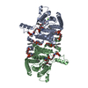 9b4eMC 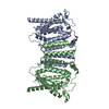 9b4fC 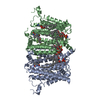 9b4gC M: atomic model generated by this map C: citing same article ( |
|---|---|
| Similar structure data | Similarity search - Function & homology  F&H Search F&H Search |
- Links
Links
| EMDB pages |  EMDB (EBI/PDBe) / EMDB (EBI/PDBe) /  EMDataResource EMDataResource |
|---|
- Map
Map
| File |  Download / File: emd_44178.map.gz / Format: CCP4 / Size: 103 MB / Type: IMAGE STORED AS FLOATING POINT NUMBER (4 BYTES) Download / File: emd_44178.map.gz / Format: CCP4 / Size: 103 MB / Type: IMAGE STORED AS FLOATING POINT NUMBER (4 BYTES) | ||||||||||||||||||||||||||||||||||||
|---|---|---|---|---|---|---|---|---|---|---|---|---|---|---|---|---|---|---|---|---|---|---|---|---|---|---|---|---|---|---|---|---|---|---|---|---|---|
| Projections & slices | Image control
Images are generated by Spider. | ||||||||||||||||||||||||||||||||||||
| Voxel size | X=Y=Z: 0.8343 Å | ||||||||||||||||||||||||||||||||||||
| Density |
| ||||||||||||||||||||||||||||||||||||
| Symmetry | Space group: 1 | ||||||||||||||||||||||||||||||||||||
| Details | EMDB XML:
|
-Supplemental data
-Half map: #2
| File | emd_44178_half_map_1.map | ||||||||||||
|---|---|---|---|---|---|---|---|---|---|---|---|---|---|
| Projections & Slices |
| ||||||||||||
| Density Histograms |
-Half map: #1
| File | emd_44178_half_map_2.map | ||||||||||||
|---|---|---|---|---|---|---|---|---|---|---|---|---|---|
| Projections & Slices |
| ||||||||||||
| Density Histograms |
- Sample components
-Entire : PSS1
| Entire | Name: PSS1 |
|---|---|
| Components |
|
-Supramolecule #1: PSS1
| Supramolecule | Name: PSS1 / type: complex / ID: 1 / Parent: 0 / Macromolecule list: #1 |
|---|---|
| Source (natural) | Organism:  Homo sapiens (human) Homo sapiens (human) |
-Macromolecule #1: Phosphatidylserine synthase 1
| Macromolecule | Name: Phosphatidylserine synthase 1 / type: protein_or_peptide / ID: 1 / Number of copies: 2 / Enantiomer: LEVO EC number: L-serine-phosphatidylethanolamine phosphatidyltransferase |
|---|---|
| Source (natural) | Organism:  Homo sapiens (human) Homo sapiens (human) |
| Molecular weight | Theoretical: 48.359551 KDa |
| Recombinant expression | Organism:  Homo sapiens (human) Homo sapiens (human) |
| Sequence | String: MASCVGSRTL SKDDVNYKMH FRMINEQQVE DITIDFFYRP HTITLLSFTI VSLMYFAFTR DDSVPEDNIW RGILSVIFFF LIISVLAFP NGPFTRPHPA LWRMVFGLSV LYFLFLVFLL FLNFEQVKSL MYWLDPNLRY ATREADVMEY AVNCHVITWE R IISHFDIF ...String: MASCVGSRTL SKDDVNYKMH FRMINEQQVE DITIDFFYRP HTITLLSFTI VSLMYFAFTR DDSVPEDNIW RGILSVIFFF LIISVLAFP NGPFTRPHPA LWRMVFGLSV LYFLFLVFLL FLNFEQVKSL MYWLDPNLRY ATREADVMEY AVNCHVITWE R IISHFDIF AFGHFWGWAM KALLIRSYGL CWTISITWEL TELFFMHLLP NFAECWWDQV ILDILLCNGG GIWLGMVVCR FL EMRTYHW ASFKDIHTTT GKIKRAVLQF TPASWTYVRW FDPKSSFQRV AGVYLFMIIW QLTELNTFFL KHIFVFQASH PLS WGRILF IGGITAPTVR QYYAYLTDTQ CKRVGTQCWV FGVIGFLEAI VCIKFGQDLF SKTQILYVVL WLLCVAFTTF LCLY GMIWY AEHY UniProtKB: Phosphatidylserine synthase 1 |
-Macromolecule #2: O-[(R)-{[(2R)-2,3-bis(octadecanoyloxy)propyl]oxy}(hydroxy)phospho...
| Macromolecule | Name: O-[(R)-{[(2R)-2,3-bis(octadecanoyloxy)propyl]oxy}(hydroxy)phosphoryl]-L-serine type: ligand / ID: 2 / Number of copies: 4 / Formula: P5S |
|---|---|
| Molecular weight | Theoretical: 792.075 Da |
| Chemical component information | 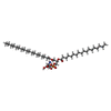 ChemComp-P5S: |
-Macromolecule #3: 1-palmitoyl-2-oleoyl-sn-glycero-3-phosphocholine
| Macromolecule | Name: 1-palmitoyl-2-oleoyl-sn-glycero-3-phosphocholine / type: ligand / ID: 3 / Number of copies: 4 / Formula: LBN |
|---|---|
| Molecular weight | Theoretical: 760.076 Da |
| Chemical component information | 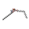 ChemComp-LBN: |
-Macromolecule #4: (2R)-3-{[(S)-hydroxy{[(1R,2R,3R,4R,5R,6S)-2,3,4,5,6-pentahydroxyc...
| Macromolecule | Name: (2R)-3-{[(S)-hydroxy{[(1R,2R,3R,4R,5R,6S)-2,3,4,5,6-pentahydroxycyclohexyl]oxy}phosphoryl]oxy}propane-1,2-diyl dihexadecanoate type: ligand / ID: 4 / Number of copies: 2 / Formula: A1AIS |
|---|---|
| Molecular weight | Theoretical: 811.032 Da |
-Macromolecule #5: [(2~{R})-1-[2-azanylethoxy(oxidanyl)phosphoryl]oxy-3-hexadecanoyl...
| Macromolecule | Name: [(2~{R})-1-[2-azanylethoxy(oxidanyl)phosphoryl]oxy-3-hexadecanoyloxy-propan-2-yl] (~{Z})-octadec-9-enoate type: ligand / ID: 5 / Number of copies: 2 / Formula: 6OU |
|---|---|
| Molecular weight | Theoretical: 717.996 Da |
| Chemical component information | 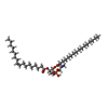 ChemComp-6OU: |
-Macromolecule #6: CALCIUM ION
| Macromolecule | Name: CALCIUM ION / type: ligand / ID: 6 / Number of copies: 2 / Formula: CA |
|---|---|
| Molecular weight | Theoretical: 40.078 Da |
-Macromolecule #7: water
| Macromolecule | Name: water / type: ligand / ID: 7 / Number of copies: 16 / Formula: HOH |
|---|---|
| Molecular weight | Theoretical: 18.015 Da |
| Chemical component information |  ChemComp-HOH: |
-Experimental details
-Structure determination
| Method | cryo EM |
|---|---|
 Processing Processing | single particle reconstruction |
| Aggregation state | particle |
- Sample preparation
| Buffer | pH: 7.5 |
|---|---|
| Vitrification | Cryogen name: ETHANE |
- Electron microscopy
| Microscope | FEI TITAN KRIOS |
|---|---|
| Image recording | Film or detector model: GATAN K3 (6k x 4k) / Average electron dose: 60.0 e/Å2 |
| Electron beam | Acceleration voltage: 300 kV / Electron source:  FIELD EMISSION GUN FIELD EMISSION GUN |
| Electron optics | Illumination mode: FLOOD BEAM / Imaging mode: BRIGHT FIELD / Nominal defocus max: 1.8 µm / Nominal defocus min: 0.8 µm |
| Experimental equipment |  Model: Titan Krios / Image courtesy: FEI Company |
- Image processing
| Startup model | Type of model: OTHER |
|---|---|
| Final reconstruction | Resolution.type: BY AUTHOR / Resolution: 2.7 Å / Resolution method: FSC 0.143 CUT-OFF / Number images used: 384020 |
| Initial angle assignment | Type: NOT APPLICABLE |
| Final angle assignment | Type: NOT APPLICABLE |
 Movie
Movie Controller


 Z (Sec.)
 Y (Row.)
 X (Col.)


