+ Open data
- Basic information
| Entry | Database: PDB / ID: 9b4f | ||||||
|---|---|---|---|---|---|---|---|
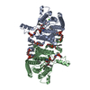
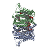
| Title | Structure of human PSS1-P269S | ||||||
 Components Components | Phosphatidylserine synthase 1 | ||||||
 Keywords Keywords | MEMBRANE PROTEIN | ||||||
| Function / homology |  Function and homology information Function and homology informationL-serine-phosphatidylcholine phosphatidyltransferase activity / L-serine-phosphatidylethanolamine phosphatidyltransferase / L-serine-phosphatidylethanolamine phosphatidyltransferase activity / Synthesis of PS / phosphatidylserine biosynthetic process / transferase activity / endoplasmic reticulum membrane / membrane Similarity search - Function | ||||||
| Biological species |  Homo sapiens (human) Homo sapiens (human) | ||||||
| Method | ELECTRON MICROSCOPY / single particle reconstruction / cryo EM / Resolution: 3.27 Å | ||||||
 Authors Authors | Long, T. / Li, X. | ||||||
| Funding support |  United States, 1items United States, 1items
| ||||||
 Citation Citation |  Journal: Cell / Year: 2024 Journal: Cell / Year: 2024Title: Molecular insights into human phosphatidylserine synthase 1 reveal its inhibition promotes LDL uptake. Authors: Tao Long / Dongyu Li / Goncalo Vale / Yaoyukun Jiang / Philip Schmiege / Zhongyue J Yang / Jeffrey G McDonald / Xiaochun Li /  Abstract: In mammalian cells, two phosphatidylserine (PS) synthases drive PS synthesis. Gain-of-function mutations in the Ptdss1 gene lead to heightened PS production, causing Lenz-Majewski syndrome (LMS). ...In mammalian cells, two phosphatidylserine (PS) synthases drive PS synthesis. Gain-of-function mutations in the Ptdss1 gene lead to heightened PS production, causing Lenz-Majewski syndrome (LMS). Recently, pharmacological inhibition of PSS1 has been shown to suppress tumorigenesis. Here, we report the cryo-EM structures of wild-type human PSS1 (PSS1), the LMS-causing Pro269Ser mutant (PSS1), and PSS1 in complex with its inhibitor DS55980254. PSS1 contains 10 transmembrane helices (TMs), with TMs 4-8 forming a catalytic core in the luminal leaflet. These structures revealed a working mechanism of PSS1 akin to the postulated mechanisms of the membrane-bound O-acyltransferase family. Additionally, we showed that both PS and DS55980254 can allosterically inhibit PSS1 and that inhibition by DS55980254 activates the SREBP pathways, thus enhancing the expression of LDL receptors and increasing cellular LDL uptake. This work uncovers a mechanism of mammalian PS synthesis and suggests that selective PSS1 inhibitors have the potential to lower blood cholesterol levels. | ||||||
| History |
|
- Structure visualization
| Structure viewer | Molecule:  Molmil Molmil Jmol/JSmol Jmol/JSmol |
|---|
- Downloads & links
- Download
| PDBx/mmCIF format |  9b4f.cif.gz 9b4f.cif.gz | 135.6 KB | Display |  PDBx/mmCIF format PDBx/mmCIF format |
|---|---|---|---|---|
| PDB format |  pdb9b4f.ent.gz pdb9b4f.ent.gz | 104.5 KB | Display |  PDB format PDB format |
| PDBx/mmJSON format |  9b4f.json.gz 9b4f.json.gz | Tree view |  PDBx/mmJSON format PDBx/mmJSON format | |
| Others |  Other downloads Other downloads |
-Validation report
| Arichive directory |  https://data.pdbj.org/pub/pdb/validation_reports/b4/9b4f https://data.pdbj.org/pub/pdb/validation_reports/b4/9b4f ftp://data.pdbj.org/pub/pdb/validation_reports/b4/9b4f ftp://data.pdbj.org/pub/pdb/validation_reports/b4/9b4f | HTTPS FTP |
|---|
-Related structure data
| Related structure data |  44179MC  9b4eC  9b4gC M: map data used to model this data C: citing same article ( |
|---|---|
| Similar structure data | Similarity search - Function & homology  F&H Search F&H Search |
- Links
Links
- Assembly
| Deposited unit | 
|
|---|---|
| 1 |
|
- Components
| #1: Protein | Mass: 56577.312 Da / Num. of mol.: 2 / Mutation: P269S Source method: isolated from a genetically manipulated source Source: (gene. exp.)  Homo sapiens (human) / Gene: PTDSS1, KIAA0024, PSSA / Production host: Homo sapiens (human) / Gene: PTDSS1, KIAA0024, PSSA / Production host:  Homo sapiens (human) Homo sapiens (human)References: UniProt: P48651, L-serine-phosphatidylethanolamine phosphatidyltransferase #2: Chemical | Has ligand of interest | N | Has protein modification | Y | |
|---|
-Experimental details
-Experiment
| Experiment | Method: ELECTRON MICROSCOPY |
|---|---|
| EM experiment | Aggregation state: PARTICLE / 3D reconstruction method: single particle reconstruction |
- Sample preparation
| Component | Name: Human PSS1-P269S / Type: COMPLEX / Entity ID: #1 / Source: RECOMBINANT |
|---|---|
| Source (natural) | Organism:  Homo sapiens (human) Homo sapiens (human) |
| Source (recombinant) | Organism:  Homo sapiens (human) Homo sapiens (human) |
| Buffer solution | pH: 7.5 |
| Specimen | Embedding applied: NO / Shadowing applied: NO / Staining applied: NO / Vitrification applied: YES |
| Vitrification | Cryogen name: ETHANE |
- Electron microscopy imaging
| Experimental equipment |  Model: Titan Krios / Image courtesy: FEI Company |
|---|---|
| Microscopy | Model: FEI TITAN KRIOS |
| Electron gun | Electron source:  FIELD EMISSION GUN / Accelerating voltage: 300 kV / Illumination mode: FLOOD BEAM FIELD EMISSION GUN / Accelerating voltage: 300 kV / Illumination mode: FLOOD BEAM |
| Electron lens | Mode: BRIGHT FIELD / Nominal defocus max: 1800 nm / Nominal defocus min: 800 nm |
| Image recording | Electron dose: 60 e/Å2 / Film or detector model: FEI FALCON IV (4k x 4k) |
- Processing
| EM software | Name: PHENIX / Version: 1.17_3644: / Category: model refinement | ||||||||||||||||||||||||
|---|---|---|---|---|---|---|---|---|---|---|---|---|---|---|---|---|---|---|---|---|---|---|---|---|---|
| CTF correction | Type: NONE | ||||||||||||||||||||||||
| 3D reconstruction | Resolution: 3.27 Å / Resolution method: FSC 0.143 CUT-OFF / Num. of particles: 155373 / Symmetry type: POINT | ||||||||||||||||||||||||
| Refine LS restraints |
|
 Movie
Movie Controller


 PDBj
PDBj

