 Movie
Movie Controller

[English] 日本語
 Yorodumi
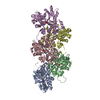
- EMDB-42918: Cryo-EM Structure of Wildtype Smooth Muscle Gamma Actin (ACTG2) -
+ Open data

- Basic information
| Entry |  | |||||||||
|---|---|---|---|---|---|---|---|---|---|---|
| Title | Cryo-EM Structure of Wildtype Smooth Muscle Gamma Actin (ACTG2) | |||||||||
 Map data Map data | ||||||||||
 Sample Sample |
| |||||||||
 Keywords Keywords | Filament / Actin / Smooth Muscle / CYTOSOLIC PROTEIN / STRUCTURAL PROTEIN | |||||||||
| Function / homology |  Function and homology information Function and homology informationmyosin filament / mesenchyme migration / Smooth Muscle Contraction / filopodium / cell periphery / Hydrolases; Acting on acid anhydrides; Acting on acid anhydrides to facilitate cellular and subcellular movement / lamellipodium / cell body / cytoskeleton / blood microparticle ...myosin filament / mesenchyme migration / Smooth Muscle Contraction / filopodium / cell periphery / Hydrolases; Acting on acid anhydrides; Acting on acid anhydrides to facilitate cellular and subcellular movement / lamellipodium / cell body / cytoskeleton / blood microparticle / hydrolase activity / positive regulation of gene expression / extracellular space / extracellular exosome / ATP binding / cytoplasm / cytosol Similarity search - Function | |||||||||
| Biological species |  Homo sapiens (human) Homo sapiens (human) | |||||||||
| Method | helical reconstruction / cryo EM / Resolution: 2.45 Å | |||||||||
 Authors Authors | Palmer NJ / Carman PJ / Ceron RH / Dominguez R | |||||||||
| Funding support |  United States, 1 items United States, 1 items
| |||||||||
 Citation Citation | Journal: Sci Adv / Year: 2024 Title: Molecular mechanisms linking missense ACTG2 mutations to visceral myopathy. Authors: Rachel H Ceron / Faviolla A Báez-Cruz / Nicholas J Palmer / Peter J Carman / Malgorzata Boczkowska / Robert O Heuckeroth / E Michael Ostap / Roberto Dominguez / Abstract: Visceral myopathy is a life-threatening disease characterized by muscle weakness in the bowel, bladder, and uterus. Mutations in smooth muscle γ-actin (ACTG2) are the most common cause of the ...Visceral myopathy is a life-threatening disease characterized by muscle weakness in the bowel, bladder, and uterus. Mutations in smooth muscle γ-actin (ACTG2) are the most common cause of the disease, but the mechanisms by which the mutations alter muscle function are unknown. Here, we examined four prevalent ACTG2 mutations (R40C, R148C, R178C, and R257C) that cause different disease severity and are spread throughout the actin fold. R178C displayed premature degradation, R148C disrupted interactions with actin-binding proteins, R40C inhibited polymerization, and R257C destabilized filaments. Because these mutations are heterozygous, we also analyzed 50/50 mixtures with wild-type (WT) ACTG2. The WT/R40C mixture impaired filament nucleation by leiomodin 1, and WT/R257C produced filaments that were easily fragmented by smooth muscle myosin. Smooth muscle tropomyosin isoform Tpm1.4 partially rescued the defects of R40C and R257C. Cryo-electron microscopy structures of filaments formed by R40C and R257C revealed disrupted intersubunit contacts. The biochemical and structural properties of the mutants correlate with their genotype-specific disease severity. | |||||||||
| History |
|
- Structure visualization
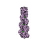
| Supplemental images |
|---|

- Downloads & links
-EMDB archive
| Map data | emd_42918.map.gz | 32.5 MB | EMDB map data format | |
|---|---|---|---|---|
| Header (meta data) | emd-42918-v30.xmlemd-42918.xml | 18.6 KB 18.6 KB | Display Display | EMDB header |
| FSC (resolution estimation) | emd_42918_fsc.xml | 8.4 KB | Display | FSC data file |
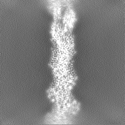
| Images | 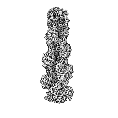 emd_42918.png emd_42918.png | 71.2 KB | ||
| Masks | emd_42918_msk_1.map | 64 MB | Mask map | |
| Filedesc metadata | emd-42918.cif.gz | 6.5 KB | ||
| Others | emd_42918_half_map_1.map.gzemd_42918_half_map_2.map.gz | 59.4 MB 59.4 MB | ||
| Archive directory |  http://ftp.pdbj.org/pub/emdb/structures/EMD-42918ftp://ftp.pdbj.org/pub/emdb/structures/EMD-42918 http://ftp.pdbj.org/pub/emdb/structures/EMD-42918ftp://ftp.pdbj.org/pub/emdb/structures/EMD-42918 | HTTPS FTP |
-Validation report
| Summary document | emd_42918_validation.pdf.gz | 940.5 KB | Display | EMDB validaton report |
|---|---|---|---|---|
| Full document | emd_42918_full_validation.pdf.gz | 940 KB | Display | |
| Data in XML | emd_42918_validation.xml.gz | 16.4 KB | Display | |
| Data in CIF | emd_42918_validation.cif.gz | 21.1 KB | Display | |
| Arichive directory | https://ftp.pdbj.org/pub/emdb/validation_reports/EMD-42918ftp://ftp.pdbj.org/pub/emdb/validation_reports/EMD-42918 | HTTPS FTP |
-Related structure data
| Related structure data |  8v2oMC 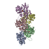 8v30C M: atomic model generated by this map C: citing same article ( |
|---|---|
| Similar structure data |
-Links
| EMDB pages | EMDB (EBI/PDBe) / EMDataResource |
|---|---|
| Related items in Molecule of the Month |


-Map
| File | Download / File: emd_42918.map.gz / Format: CCP4 / Size: 64 MB / Type: IMAGE STORED AS FLOATING POINT NUMBER (4 BYTES) | ||||||||||||||||||||
|---|---|---|---|---|---|---|---|---|---|---|---|---|---|---|---|---|---|---|---|---|---|
| Voxel size | X=Y=Z: 1.07 Å | ||||||||||||||||||||
| Density |
| ||||||||||||||||||||
| Symmetry | Space group: 1 | ||||||||||||||||||||
| Details | EMDB XML:
|
-Supplemental data
-Mask #1
| File | emd_42918_msk_1.map | ||||||||||||
|---|---|---|---|---|---|---|---|---|---|---|---|---|---|
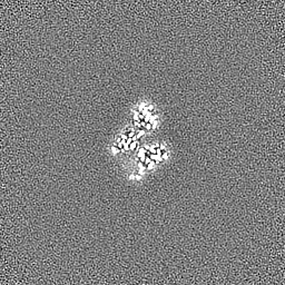
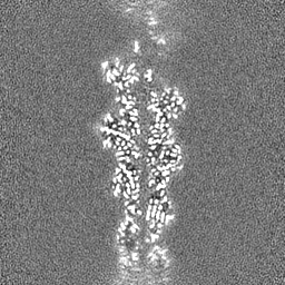
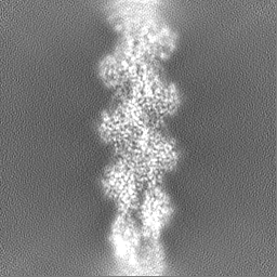
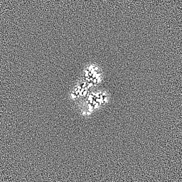
| Projections & Slices |
| ||||||||||||
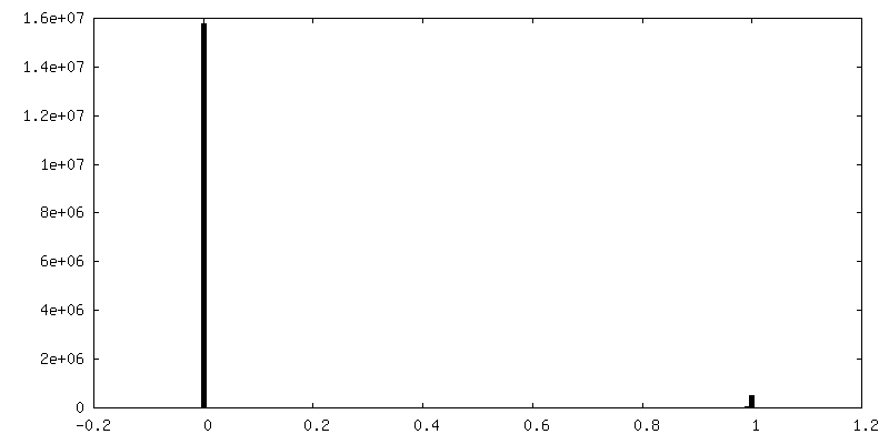
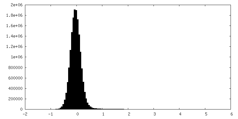
| Density Histograms |
 Z
Z Y
Y X
X


-Half map: #1
| File | emd_42918_half_map_1.map | ||||||||||||
|---|---|---|---|---|---|---|---|---|---|---|---|---|---|
| Projections & Slices |
| ||||||||||||
| Density Histograms |


-Half map: #2
| File | emd_42918_half_map_2.map | ||||||||||||
|---|---|---|---|---|---|---|---|---|---|---|---|---|---|
| Projections & Slices |
| ||||||||||||
| Density Histograms |


- Sample components
-Entire : ACTG2 filament
| Entire | Name: ACTG2 filament |
|---|---|
| Components |
|
-Supramolecule #1: ACTG2 filament
| Supramolecule | Name: ACTG2 filament / type: complex / ID: 1 / Parent: 0 / Macromolecule list: #1 / Details: ACTG2 filaments, purified from Expi293 cells |
|---|---|
| Source (natural) | Organism: Homo sapiens (human) / Tissue: Smooth |
-Macromolecule #1: Actin, gamma-enteric smooth muscle
| Macromolecule | Name: Actin, gamma-enteric smooth muscle / type: protein_or_peptide / ID: 1 / Number of copies: 5 / Enantiomer: LEVO EC number: Hydrolases; Acting on acid anhydrides; Acting on acid anhydrides to facilitate cellular and subcellular movement |
|---|---|
| Source (natural) | Organism: Homo sapiens (human) / Tissue: Smooth Muscle |
| Molecular weight | Theoretical: 41.935816 KDa |
| Recombinant expression | Organism: Homo sapiens (human) |
| Sequence | String: MCEEETTALV CDNGSGLCKA GFAGDDAPRA VFPSIVGRPR HQGVMVGMGQ KDSYVGDEAQ SKRGILTLKY PIE(HIC)GI ITN WDDMEKIWHH SFYNELRVAP EEHPTLLTEA PLNPKANREK MTQIMFETFN VPAMYVAIQA VLSLYASGRT TGIVLDS GD GVTHNVPIYE ...String: MCEEETTALV CDNGSGLCKA GFAGDDAPRA VFPSIVGRPR HQGVMVGMGQ KDSYVGDEAQ SKRGILTLKY PIE(HIC)GI ITN WDDMEKIWHH SFYNELRVAP EEHPTLLTEA PLNPKANREK MTQIMFETFN VPAMYVAIQA VLSLYASGRT TGIVLDS GD GVTHNVPIYE GYALPHAIMR LDLAGRDLTD YLMKILTERG YSFVTTAERE IVRDIKEKLC YVALDFENEM ATAASSSS L EKSYELPDGQ VITIGNERFR CPETLFQPSF IGMESAGIHE TTYNSIMKCD IDIRKDLYAN NVLSGGTTMY PGIADRMQK EITALAPSTM KIKIIAPPER KYSVWIGGSI LASLSTFQQM WISKPEYDEA GPSIVHRKCF UniProtKB: Actin, gamma-enteric smooth muscle |
-Macromolecule #2: ADENOSINE-5'-DIPHOSPHATE
| Macromolecule | Name: ADENOSINE-5'-DIPHOSPHATE / type: ligand / ID: 2 / Number of copies: 5 / Formula: ADP |
|---|---|
| Molecular weight | Theoretical: 427.201 Da |
| Chemical component information |  ChemComp-ADP: |
-Macromolecule #3: MAGNESIUM ION
| Macromolecule | Name: MAGNESIUM ION / type: ligand / ID: 3 / Number of copies: 5 / Formula: MG |
|---|---|
| Molecular weight | Theoretical: 24.305 Da |
-Experimental details
-Structure determination
| Method | cryo EM |
|---|---|
 Processing Processing | helical reconstruction |
| Aggregation state | filament |
-Sample preparation
| Concentration | 0.168 mg/mL | |||||||||||||||||||||
|---|---|---|---|---|---|---|---|---|---|---|---|---|---|---|---|---|---|---|---|---|---|---|
| Buffer | pH: 8 Component:
Details: Actin F-buffer | |||||||||||||||||||||
| Grid | Model: Quantifoil R1.2/1.3 / Material: COPPER / Mesh: 200 / Support film - Material: CARBON / Support film - topology: HOLEY / Support film - Film thickness: 12 / Pretreatment - Type: GLOW DISCHARGE / Pretreatment - Time: 120 sec. / Pretreatment - Atmosphere: AIR | |||||||||||||||||||||
| Vitrification | Cryogen name: ETHANE / Chamber humidity: 100 % / Chamber temperature: 277 K / Instrument: FEI VITROBOT MARK III / Details: Blot Force: 0 Blot Time: 2.5 s. | |||||||||||||||||||||
| Details | ACTG2 F-Actin in the ADP state. |
- Electron microscopy
| Microscope | FEI TITAN KRIOS |
|---|---|
| Image recording | Film or detector model: GATAN K3 (6k x 4k) / Number real images: 526 / Average electron dose: 45.0 e/Å2 |
| Electron beam | Acceleration voltage: 300 kV / Electron source:  FIELD EMISSION GUN FIELD EMISSION GUN |
| Electron optics | C2 aperture diameter: 100.0 µm / Illumination mode: SPOT SCAN / Imaging mode: BRIGHT FIELD / Nominal defocus max: 2.5 µm / Nominal defocus min: 0.5 µm / Nominal magnification: 81000 |
| Sample stage | Specimen holder model: FEI TITAN KRIOS AUTOGRID HOLDER / Cooling holder cryogen: NITROGEN |
| Experimental equipment |  Model: Titan Krios / Image courtesy: FEI Company |
-Image processing
| Final reconstruction | Applied symmetry - Helical parameters - Δz: 27.5 Å Applied symmetry - Helical parameters - Δ&Phi: -166.6 ° Applied symmetry - Helical parameters - Axial symmetry: C1 (asymmetric) Resolution.type: BY AUTHOR / Resolution: 2.45 Å / Resolution method: FSC 0.143 CUT-OFF / Software - Name: cryoSPARC (ver. 4.0) / Software - details: Helix Refinement / Number images used: 473627 |
|---|---|
| Startup model | Type of model: PDB ENTRY PDB model - PDB ID: Details: Individually mutated residues within the skeletal muscle actin until each subunit had the sequence of gamma smooth muscle actin. |
| Final angle assignment | Type: NOT APPLICABLE |
| FSC plot (resolution estimation) | 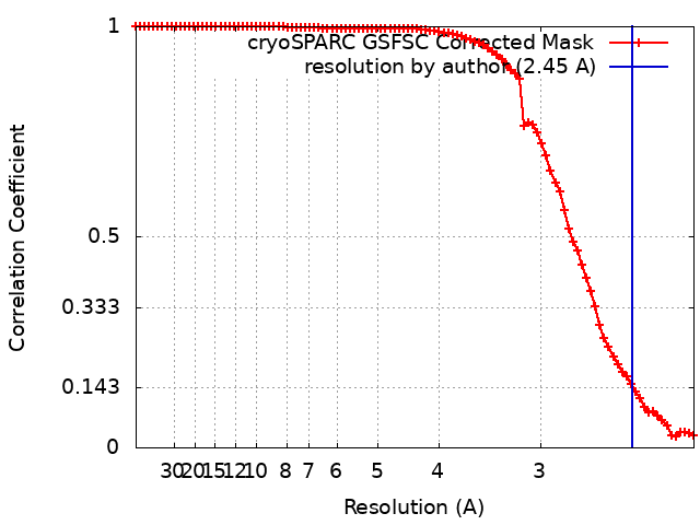 |
-Atomic model buiding 1
| Refinement | Space: REAL / Protocol: RIGID BODY FIT |
|---|---|
| Output model | PDB-8v2o: |
