Natural Sciences and Engineering Research Council (NSERC, Canada)
Canada
Citation
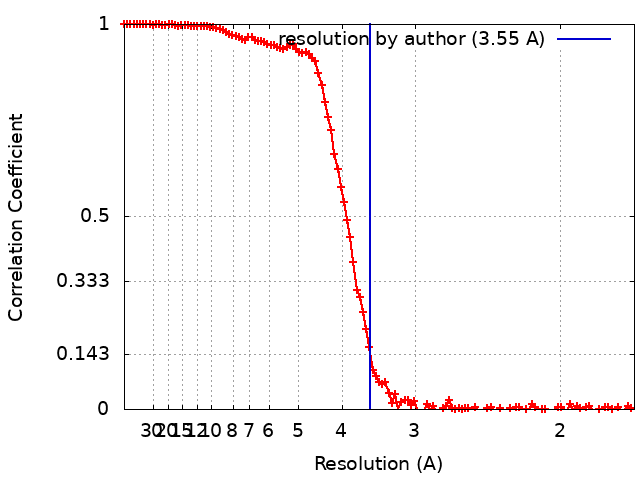
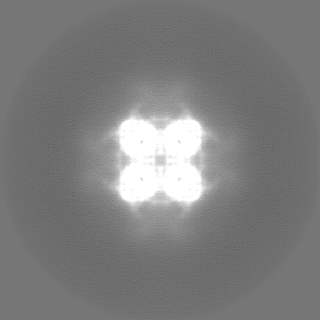
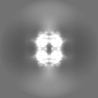
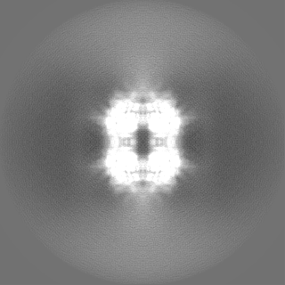
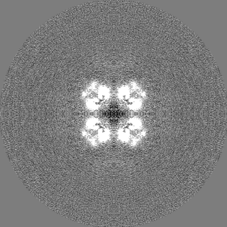
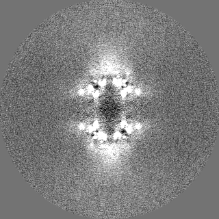
Journal: Nat Commun / Year: 2024 Title: Microfibril-associated glycoprotein 4 forms octamers that mediate interactions with elastogenic proteins and cells. Authors: Michael R Wozny / Valentin Nelea / Iram Fatima S Siddiqui / Shaynah Wanga / Vivian de Waard / Mike Strauss / Dieter P Reinhardt / Abstract: Microfibril-associated glycoprotein 4 (MFAP4) is a 36-kDa extracellular matrix glycoprotein with critical roles in organ fibrosis, chronic obstructive pulmonary disease, and cardiovascular ...Microfibril-associated glycoprotein 4 (MFAP4) is a 36-kDa extracellular matrix glycoprotein with critical roles in organ fibrosis, chronic obstructive pulmonary disease, and cardiovascular disorders, including aortic aneurysms. MFAP4 multimerises and interacts with elastogenic proteins, including fibrillin-1 and tropoelastin, and with cells via integrins. Structural details of MFAP4 and its potential interfaces for these interactions are unknown. Here, we present a cryo-electron microscopy structure of human MFAP4. In the presence of calcium, MFAP4 assembles as an octamer, where two sets of homodimers constitute the top and bottom halves of each octamer. Each homodimer is linked together by an intermolecular disulphide bond. A C34S missense mutation prevents disulphide-bond formation between monomers but does not prevent octamer assembly. The atomic model, built into the 3.55 Å cryo-EM map, suggests that salt-bridge interactions mediate homodimer assembly, while non-polar residues form the interface between octamer halves. In the absence of calcium, an MFAP4 octamer dissociates into two tetramers. Binding studies with fibrillin-1, tropoelastin, LTBP4, and small fibulins show that MFAP4 has multiple surfaces for protein-protein interactions, most of which depend upon MFAP4 octamer assembly. The C34S mutation does not affect these protein interactions or cell interactions. MFAP4 assemblies with fibrillin-1 abrogate MFAP4 interactions with cells.
In the structure databanks used in Yorodumi, some data are registered as the other names, "COVID-19 virus" and "2019-nCoV". Here are the details of the virus and the list of structure data.
Jan 31, 2019. EMDB accession codes are about to change! (news from PDBe EMDB page)
EMDB accession codes are about to change! (news from PDBe EMDB page)
The allocation of 4 digits for EMDB accession codes will soon come to an end. Whilst these codes will remain in use, new EMDB accession codes will include an additional digit and will expand incrementally as the available range of codes is exhausted. The current 4-digit format prefixed with “EMD-” (i.e. EMD-XXXX) will advance to a 5-digit format (i.e. EMD-XXXXX), and so on. It is currently estimated that the 4-digit codes will be depleted around Spring 2019, at which point the 5-digit format will come into force.
The EM Navigator/Yorodumi systems omit the EMD- prefix.
Related info.:Q: What is EMD? / ID/Accession-code notation in Yorodumi/EM Navigator
Yorodumi is a browser for structure data from EMDB, PDB, SASBDB, etc.
This page is also the successor to EM Navigator detail page, and also detail information page/front-end page for Omokage search.
The word "yorodu" (or yorozu) is an old Japanese word meaning "ten thousand". "mi" (miru) is to see.
Related info.:EMDB / PDB / SASBDB / Comparison of 3 databanks / Yorodumi Search / Aug 31, 2016. New EM Navigator & Yorodumi / Yorodumi Papers / Jmol/JSmol / Function and homology information / Changes in new EM Navigator and Yorodumi
 Movie
Movie Controller


 Open data
 Basic information
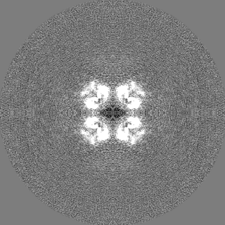
 Map data
 Sample
 Keywords
 Function and homology information
 Homo sapiens (human)
 Authors
 Canada, 2 items
  Citation
 Structure visualization
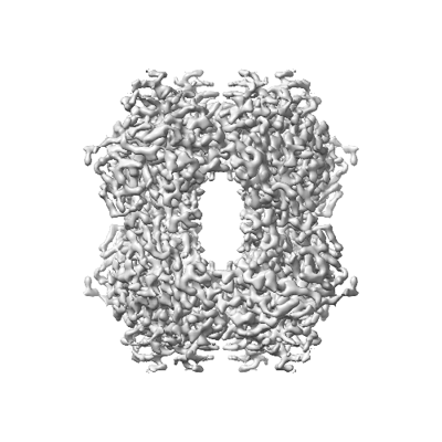
 Downloads & links
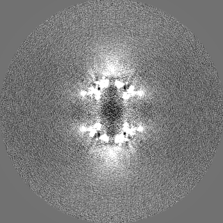
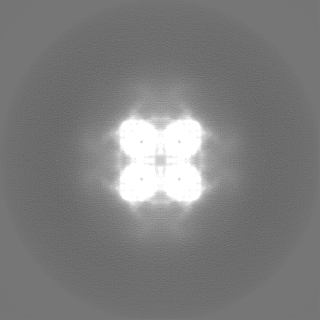
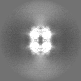
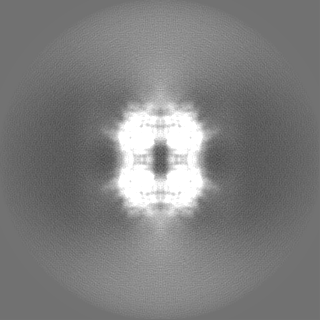
 emd_42394.png
 http://ftp.pdbj.org/pub/emdb/structures/EMD-42394


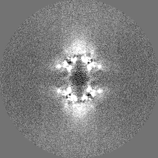
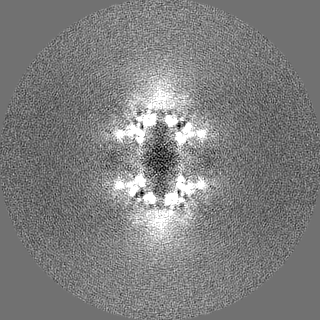
 Z (Sec.)
 Y (Row.)
 X (Col.)


 Sample components
 Processing
 Electron microscopy
 FIELD EMISSION GUN