 Movie
Movie Controller


+ Open data

- Basic information
| Entry |  | |||||||||
|---|---|---|---|---|---|---|---|---|---|---|
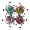
| Title | MFAP4 after treatment with EDTA/without Ca2+ | |||||||||
 Map data Map data | D2 Relion4 reconstruction after postprocessing within Relion4 of MFAP4 after EDTA treatment. | |||||||||
 Sample Sample |
| |||||||||
 Keywords Keywords | MFAP4 Octamer Extracellular Matrix / CELL ADHESION | |||||||||
| Biological species |  Homo sapiens (human) Homo sapiens (human) | |||||||||
| Method | single particle reconstruction / cryo EM / Resolution: 7.3 Å | |||||||||
 Authors Authors | Wozny MR / Nelea V / Siddiqui IFS / Wanga S / de Waard V / Strauss M / Reinhardt DP | |||||||||
| Funding support |  Canada, 2 items Canada, 2 items
| |||||||||
 Citation Citation | Journal: Nat Commun / Year: 2024 Title: Microfibril-associated glycoprotein 4 forms octamers that mediate interactions with elastogenic proteins and cells. Authors: Michael R Wozny / Valentin Nelea / Iram Fatima S Siddiqui / Shaynah Wanga / Vivian de Waard / Mike Strauss / Dieter P Reinhardt /  Abstract: Microfibril-associated glycoprotein 4 (MFAP4) is a 36-kDa extracellular matrix glycoprotein with critical roles in organ fibrosis, chronic obstructive pulmonary disease, and cardiovascular ...Microfibril-associated glycoprotein 4 (MFAP4) is a 36-kDa extracellular matrix glycoprotein with critical roles in organ fibrosis, chronic obstructive pulmonary disease, and cardiovascular disorders, including aortic aneurysms. MFAP4 multimerises and interacts with elastogenic proteins, including fibrillin-1 and tropoelastin, and with cells via integrins. Structural details of MFAP4 and its potential interfaces for these interactions are unknown. Here, we present a cryo-electron microscopy structure of human MFAP4. In the presence of calcium, MFAP4 assembles as an octamer, where two sets of homodimers constitute the top and bottom halves of each octamer. Each homodimer is linked together by an intermolecular disulphide bond. A C34S missense mutation prevents disulphide-bond formation between monomers but does not prevent octamer assembly. The atomic model, built into the 3.55 Å cryo-EM map, suggests that salt-bridge interactions mediate homodimer assembly, while non-polar residues form the interface between octamer halves. In the absence of calcium, an MFAP4 octamer dissociates into two tetramers. Binding studies with fibrillin-1, tropoelastin, LTBP4, and small fibulins show that MFAP4 has multiple surfaces for protein-protein interactions, most of which depend upon MFAP4 octamer assembly. The C34S mutation does not affect these protein interactions or cell interactions. MFAP4 assemblies with fibrillin-1 abrogate MFAP4 interactions with cells. | |||||||||
| History |
|
- Structure visualization
| Supplemental images |
|---|

- Downloads & links
-EMDB archive
| Map data | emd_42398.map.gz | 14.3 MB |  EMDB map data format EMDB map data format | |
|---|---|---|---|---|
| Header (meta data) | emd-42398-v30.xmlemd-42398.xml | 13.5 KB 13.5 KB | Display Display | EMDB header |
| FSC (resolution estimation) | emd_42398_fsc.xml | 7.4 KB | Display | FSC data file |
| Images |  emd_42398.png emd_42398.png | 25.9 KB | ||
| Filedesc metadata | emd-42398.cif.gz | 4.7 KB | ||
| Others | emd_42398_half_map_1.map.gzemd_42398_half_map_2.map.gz | 11.1 MB 11.1 MB | ||
| Archive directory |  http://ftp.pdbj.org/pub/emdb/structures/EMD-42398ftp://ftp.pdbj.org/pub/emdb/structures/EMD-42398 http://ftp.pdbj.org/pub/emdb/structures/EMD-42398ftp://ftp.pdbj.org/pub/emdb/structures/EMD-42398 | HTTPS FTP |
-Related structure data

-Links
| EMDB pages | EMDB (EBI/PDBe) / EMDataResource |
|---|
-Map
| File | Download / File: emd_42398.map.gz / Format: CCP4 / Size: 15.6 MB / Type: IMAGE STORED AS FLOATING POINT NUMBER (4 BYTES) | ||||||||||||||||||||||||||||||||||||
|---|---|---|---|---|---|---|---|---|---|---|---|---|---|---|---|---|---|---|---|---|---|---|---|---|---|---|---|---|---|---|---|---|---|---|---|---|---|
| Annotation | D2 Relion4 reconstruction after postprocessing within Relion4 of MFAP4 after EDTA treatment. | ||||||||||||||||||||||||||||||||||||
| Projections & slices | Image control
Images are generated by Spider. | ||||||||||||||||||||||||||||||||||||
| Voxel size | X=Y=Z: 1.171 Å | ||||||||||||||||||||||||||||||||||||
| Density |
| ||||||||||||||||||||||||||||||||||||
| Symmetry | Space group: 1 | ||||||||||||||||||||||||||||||||||||
| Details | EMDB XML:
|
 Z (Sec.)
 Y (Row.)
 X (Col.)


-Supplemental data
-Half map: Half map from D2 Relion4 refinement of MFAP4 after EDTA treatment.
| File | emd_42398_half_map_1.map | ||||||||||||
|---|---|---|---|---|---|---|---|---|---|---|---|---|---|
| Annotation | Half map from D2 Relion4 refinement of MFAP4 after EDTA treatment. | ||||||||||||
| Projections & Slices |
| ||||||||||||
| Density Histograms |


-Half map: Half map from D2 Relion4 refinement of MFAP4 after EDTA treatment.
| File | emd_42398_half_map_2.map | ||||||||||||
|---|---|---|---|---|---|---|---|---|---|---|---|---|---|
| Annotation | Half map from D2 Relion4 refinement of MFAP4 after EDTA treatment. | ||||||||||||
| Projections & Slices |
| ||||||||||||
| Density Histograms |


- Sample components
-Entire : MFAP4
| Entire | Name: MFAP4 |
|---|---|
| Components |
|
-Supramolecule #1: MFAP4
| Supramolecule | Name: MFAP4 / type: complex / ID: 1 / Parent: 0 / Macromolecule list: all Details: MFAP4 treated with EDTA and dialysed against TBS without Ca2+ |
|---|---|
| Source (natural) | Organism: Homo sapiens (human) |
| Molecular weight | Theoretical: 140 KDa |
-Macromolecule #1: MFAP4
| Macromolecule | Name: MFAP4 / type: protein_or_peptide / ID: 1 / Enantiomer: LEVO |
|---|---|
| Source (natural) | Organism: Homo sapiens (human) |
| Recombinant expression | Organism: Homo sapiens (human) |
| Sequence | String: CLQQPLDCDD IYAQGYQSDG VYLIYPSGPS VPVPVFCDMT TEGGKWTVFQ KRFNGSVSFF RGWNDYKLGF GRADGEYWLG LQNMHLLTLK QKYELRVDLE DFENNTAYAK YADFSISPNA VSAEEDGYTL FVAGFEDGGA GDSLSYHSGQ KFSTFDRDQD LFVQNCAALS ...String: CLQQPLDCDD IYAQGYQSDG VYLIYPSGPS VPVPVFCDMT TEGGKWTVFQ KRFNGSVSFF RGWNDYKLGF GRADGEYWLG LQNMHLLTLK QKYELRVDLE DFENNTAYAK YADFSISPNA VSAEEDGYTL FVAGFEDGGA GDSLSYHSGQ KFSTFDRDQD LFVQNCAALS SGAFWFRSCH FANLNGFYLG GSHLSYANGI NWAQWKGFYY SLKRTEMKIR RA |
-Experimental details
-Structure determination
| Method | cryo EM |
|---|---|
 Processing Processing | single particle reconstruction |
| Aggregation state | particle |
-Sample preparation
| Buffer | pH: 7 |
|---|---|
| Vitrification | Cryogen name: ETHANE / Chamber humidity: 90 % / Chamber temperature: 298 K |
- Electron microscopy
| Microscope | TFS KRIOS |
|---|---|
| Image recording | Film or detector model: GATAN K3 BIOQUANTUM (6k x 4k) / Average electron dose: 80.0 e/Å2 |
| Electron beam | Acceleration voltage: 300 kV / Electron source:  FIELD EMISSION GUN FIELD EMISSION GUN |
| Electron optics | Illumination mode: FLOOD BEAM / Imaging mode: BRIGHT FIELD / Nominal defocus max: 3.0 µm / Nominal defocus min: 1.5 µm |
| Experimental equipment |  Model: Titan Krios / Image courtesy: FEI Company |

