 Movie
Movie Controller


+ Open data

- Basic information
| Entry |  | |||||||||
|---|---|---|---|---|---|---|---|---|---|---|
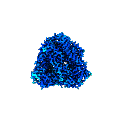
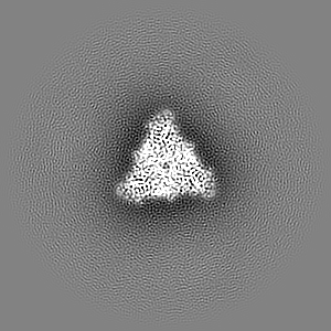
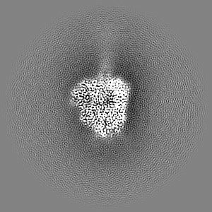
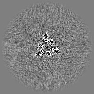
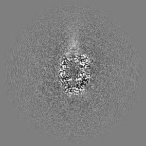
| Title | Langya henipavirus fusion protein in prefusion state | |||||||||
 Map data Map data | ||||||||||
 Sample Sample |
| |||||||||
 Keywords Keywords | Langya / henipavirus / fusion protein / LayVF / prefusion / Structural Genomics / Seattle Structural Genomics Center for Infectious Disease / SSGCID / VIRAL PROTEIN | |||||||||
| Biological species |  Langya virus Langya virus | |||||||||
| Method | single particle reconstruction / cryo EM / Resolution: 2.8 Å | |||||||||
 Authors Authors | Wang Z / Veesler D / Seattle Structural Genomics Center for Infectious Disease (SSGCID) | |||||||||
| Funding support |  United States, 1 items United States, 1 items
| |||||||||
 Citation Citation | Journal: Proc Natl Acad Sci U S A / Year: 2024 Title: Structure and design of Langya virus glycoprotein antigens. Authors: Zhaoqian Wang / Matthew McCallum / Lianying Yan / Cecily A Gibson / William Sharkey / Young-Jun Park / Ha V Dang / Moushimi Amaya / Ashley Person / Christopher C Broder / David Veesler / Abstract: Langya virus (LayV) is a recently discovered henipavirus (HNV), isolated from febrile patients in China. HNV entry into host cells is mediated by the attachment (G) and fusion (F) glycoproteins which ...Langya virus (LayV) is a recently discovered henipavirus (HNV), isolated from febrile patients in China. HNV entry into host cells is mediated by the attachment (G) and fusion (F) glycoproteins which are the main targets of neutralizing antibodies. We show here that the LayV F and G glycoproteins promote membrane fusion with human, mouse, and hamster target cells using a different, yet unknown, receptor than Nipah virus (NiV) and Hendra virus (HeV) and that NiV- and HeV-elicited monoclonal and polyclonal antibodies do not cross-react with LayV F and G. We determined cryoelectron microscopy structures of LayV F, in the prefusion and postfusion states, and of LayV G, revealing their conformational landscape and distinct antigenicity relative to NiV and HeV. We computationally designed stabilized LayV G constructs and demonstrate the generalizability of an HNV F prefusion-stabilization strategy. Our data will support the development of vaccines and therapeutics against LayV and closely related HNVs. | |||||||||
| History |
|
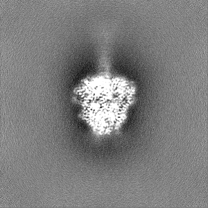
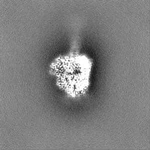
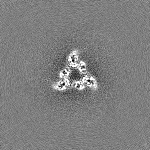
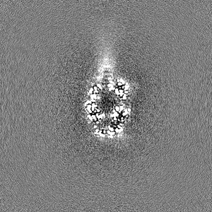
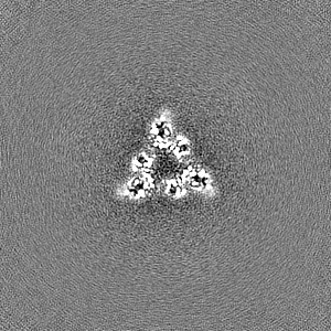
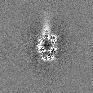
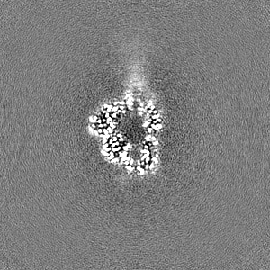
- Structure visualization
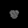
| Supplemental images |
|---|
- Downloads & links
-EMDB archive
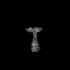
| Map data | emd_41640.map.gz | 52.1 MB |  EMDB map data format EMDB map data format | |
|---|---|---|---|---|
| Header (meta data) | emd-41640-v30.xmlemd-41640.xml | 18.6 KB 18.6 KB | Display Display | EMDB header |
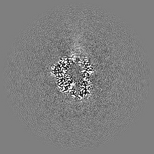
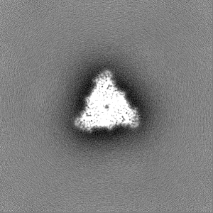
| Images |  emd_41640.png emd_41640.png | 58 KB | ||
| Filedesc metadata | emd-41640.cif.gz | 6.1 KB | ||
| Others | emd_41640_additional_1.map.gzemd_41640_half_map_1.map.gzemd_41640_half_map_2.map.gz | 97.2 MB 95.6 MB 95.6 MB | ||
| Archive directory |  http://ftp.pdbj.org/pub/emdb/structures/EMD-41640ftp://ftp.pdbj.org/pub/emdb/structures/EMD-41640 http://ftp.pdbj.org/pub/emdb/structures/EMD-41640ftp://ftp.pdbj.org/pub/emdb/structures/EMD-41640 | HTTPS FTP |
-Related structure data
| Related structure data | 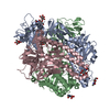 8tvfMC  8tvbC  8tveC 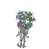 8tvgC 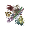 8tvhC  8tviC 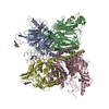 8vwpC C: citing same article ( M: atomic model generated by this map |
|---|


-Links
| EMDB pages | EMDB (EBI/PDBe) / EMDataResource |
|---|
-Map
| File | Download / File: emd_41640.map.gz / Format: CCP4 / Size: 103 MB / Type: IMAGE STORED AS FLOATING POINT NUMBER (4 BYTES) | ||||||||||||||||||||||||||||||||||||
|---|---|---|---|---|---|---|---|---|---|---|---|---|---|---|---|---|---|---|---|---|---|---|---|---|---|---|---|---|---|---|---|---|---|---|---|---|---|
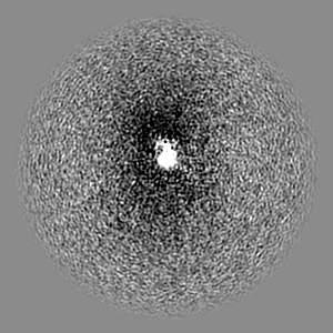
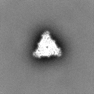
| Projections & slices | Image control
Images are generated by Spider. | ||||||||||||||||||||||||||||||||||||
| Voxel size | X=Y=Z: 1.0004 Å | ||||||||||||||||||||||||||||||||||||
| Density |
| ||||||||||||||||||||||||||||||||||||
| Symmetry | Space group: 1 | ||||||||||||||||||||||||||||||||||||
| Details | EMDB XML:
|
 Z (Sec.)
 Y (Row.)
 X (Col.)


-Supplemental data
-Additional map: Unsharpened
| File | emd_41640_additional_1.map | ||||||||||||
|---|---|---|---|---|---|---|---|---|---|---|---|---|---|
| Annotation | Unsharpened | ||||||||||||
| Projections & Slices |
| ||||||||||||
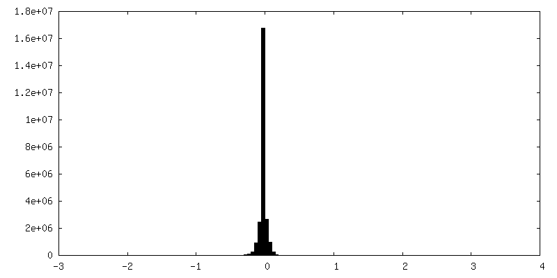
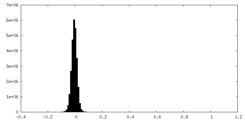
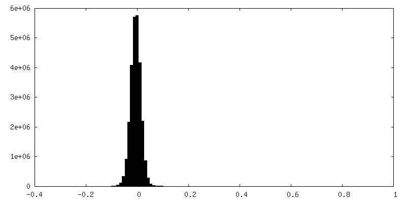
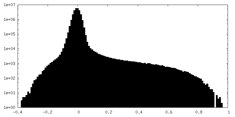
| Density Histograms |

-Half map: #1
| File | emd_41640_half_map_1.map | ||||||||||||
|---|---|---|---|---|---|---|---|---|---|---|---|---|---|
| Projections & Slices |
| ||||||||||||
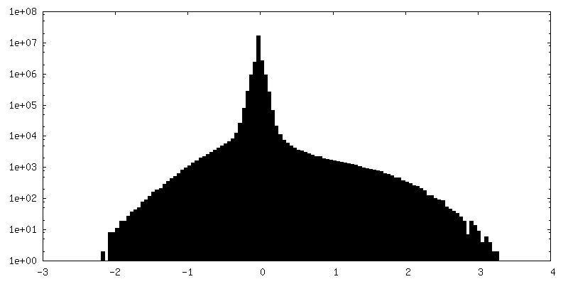
| Density Histograms |


-Half map: #2
| File | emd_41640_half_map_2.map | ||||||||||||
|---|---|---|---|---|---|---|---|---|---|---|---|---|---|
| Projections & Slices |
| ||||||||||||
| Density Histograms |
- Sample components
-Entire : Langya henipavirus fusion protein in prefusion state
| Entire | Name: Langya henipavirus fusion protein in prefusion state |
|---|---|
| Components |
|
-Supramolecule #1: Langya henipavirus fusion protein in prefusion state
| Supramolecule | Name: Langya henipavirus fusion protein in prefusion state / type: complex / ID: 1 / Parent: 0 / Macromolecule list: #1 |
|---|---|
| Source (natural) | Organism: Langya virus |
-Macromolecule #1: Langya henipavirus fusion protein in prefusion state
| Macromolecule | Name: Langya henipavirus fusion protein in prefusion state / type: protein_or_peptide / ID: 1 / Number of copies: 3 / Enantiomer: LEVO |
|---|---|
| Source (natural) | Organism: Langya virus |
| Molecular weight | Theoretical: 58.480812 KDa |
| Recombinant expression | Organism:  Homo sapiens (human) Homo sapiens (human) |
| Sequence | String: MAFLKSAIIC YLLFYPHIVK SSLHYDSLSK VGIIKGLTYN YKIKGSPSTK LMVVKLIPNI DGVRNCTQKQ FDEYKNLVKN VLEPVKLAL NAMLDNVKSG NNKYRFAGAI MAGVALGVAT AATVTAGIAL HRSNENAQAI ANMKNAIQNT NEAVKQLQLA N KQTLAVID ...String: MAFLKSAIIC YLLFYPHIVK SSLHYDSLSK VGIIKGLTYN YKIKGSPSTK LMVVKLIPNI DGVRNCTQKQ FDEYKNLVKN VLEPVKLAL NAMLDNVKSG NNKYRFAGAI MAGVALGVAT AATVTAGIAL HRSNENAQAI ANMKNAIQNT NEAVKQLQLA N KQTLAVID TIRGEINNNI IPVINQLSCD TIGLSVGIKL TQYYSEILTA FGPALQNPVN TRITIQAISS VFNRNFDELL KI MGYTSGD LYEILHSGLI RGNIIDVDVE AGYIALEIEF PNLTLVPNAV VQELMPISYN VDGDEWVTLV PRFVLTRTTL LSN IDTSRC TVTESSVICD NDYALPMSYE LIGCLQGDTS KCAREKVVSS YVPRFALSDG LVYANCLNTI CRCMDTDTPI SQSL GTTVS LLDNKKCLVY QVGDILISVG SYLGEGEYSA DNVELGPPVV IDKIDIGNQL AGINQTLQNA EDYIEKSEEF LKGIN PSMK QIEDKIEEIL SKIYHIENEI ARIKKLIGEA PGGSIEGRGS GGGSHHHHHH |
-Macromolecule #3: 2-acetamido-2-deoxy-beta-D-glucopyranose
| Macromolecule | Name: 2-acetamido-2-deoxy-beta-D-glucopyranose / type: ligand / ID: 3 / Number of copies: 3 / Formula: NAG |
|---|---|
| Molecular weight | Theoretical: 221.208 Da |
| Chemical component information |  ChemComp-NAG: |
-Macromolecule #4: water
| Macromolecule | Name: water / type: ligand / ID: 4 / Number of copies: 564 / Formula: HOH |
|---|---|
| Molecular weight | Theoretical: 18.015 Da |
| Chemical component information |  ChemComp-HOH: |
-Experimental details
-Structure determination
| Method | cryo EM |
|---|---|
 Processing Processing | single particle reconstruction |
| Aggregation state | particle |
-Sample preparation
| Buffer | pH: 8 |
|---|---|
| Grid | Model: C-flat-2/2 / Material: COPPER / Support film - Material: GRAPHENE OXIDE / Support film - topology: CONTINUOUS |
| Vitrification | Cryogen name: ETHANE / Chamber humidity: 100 % / Chamber temperature: 24 K / Instrument: FEI VITROBOT MARK IV |
- Electron microscopy
| Microscope | TFS KRIOS |
|---|---|
| Image recording | Film or detector model: GATAN K3 (6k x 4k) / Average electron dose: 63.0 e/Å2 |
| Electron beam | Acceleration voltage: 300 kV / Electron source:  FIELD EMISSION GUN FIELD EMISSION GUN |
| Electron optics | Illumination mode: FLOOD BEAM / Imaging mode: BRIGHT FIELD / Cs: 2.7 mm / Nominal defocus max: 1.7 µm / Nominal defocus min: 0.7000000000000001 µm |
| Experimental equipment |  Model: Titan Krios / Image courtesy: FEI Company |
-Image processing
| Startup model | Type of model: OTHER |
|---|---|
| Final reconstruction | Applied symmetry - Point group: C3 (3 fold cyclic) / Resolution.type: BY AUTHOR / Resolution: 2.8 Å / Resolution method: FSC 0.143 CUT-OFF / Number images used: 53917 |
| Initial angle assignment | Type: MAXIMUM LIKELIHOOD |
| Final angle assignment | Type: MAXIMUM LIKELIHOOD |
