European Union, United States, Czech Republic, 4 items
Organization
Grant number
Country
European Research Council (ERC)
Grant ERC-AdG 786714 (LIFETimeS)
European Union
Department of Energy (DOE, United States)
DE-SC0020606
United States
National Institutes of Health/National Institute of General Medical Sciences (NIH/NIGMS)
GM127018
United States
Czech Science Foundation
19-28323X
Czech Republic
Citation
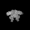
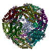
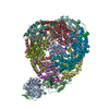
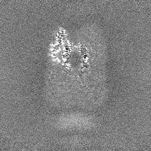
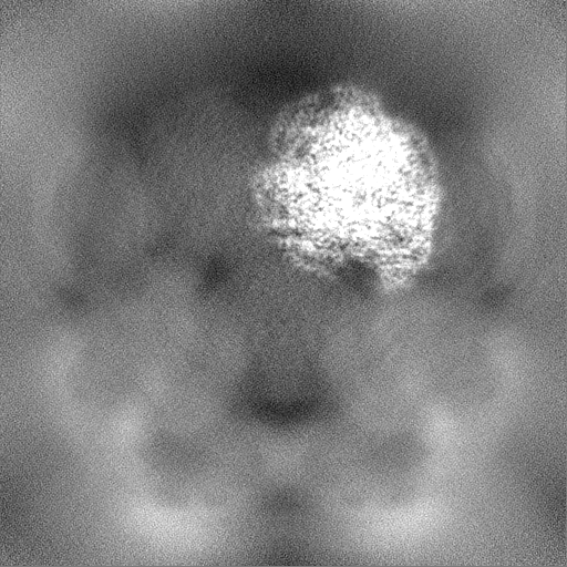
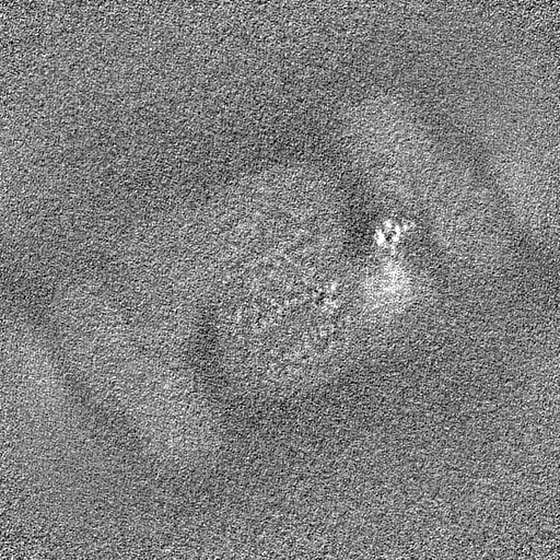
Journal: Sci Adv / Year: 2024 Title: Structural and quantum chemical basis for OCP-mediated quenching of phycobilisomes. Authors: Paul V Sauer / Lorenzo Cupellini / Markus Sutter / Mattia Bondanza / María Agustina Domínguez Martin / Henning Kirst / David Bína / Adrian Fujiet Koh / Abhay Kotecha / Basil J Greber / ...Authors: Paul V Sauer / Lorenzo Cupellini / Markus Sutter / Mattia Bondanza / María Agustina Domínguez Martin / Henning Kirst / David Bína / Adrian Fujiet Koh / Abhay Kotecha / Basil J Greber / Eva Nogales / Tomáš Polívka / Benedetta Mennucci / Cheryl A Kerfeld / Abstract: Cyanobacteria use large antenna complexes called phycobilisomes (PBSs) for light harvesting. However, intense light triggers non-photochemical quenching, where the orange carotenoid protein (OCP) ...Cyanobacteria use large antenna complexes called phycobilisomes (PBSs) for light harvesting. However, intense light triggers non-photochemical quenching, where the orange carotenoid protein (OCP) binds to PBS, dissipating excess energy as heat. The mechanism of efficiently transferring energy from phycocyanobilins in PBS to canthaxanthin in OCP remains insufficiently understood. Using cryo-electron microscopy, we unveiled the OCP-PBS complex structure at 1.6- to 2.1-angstrom resolution, showcasing its inherent flexibility. Using multiscale quantum chemistry, we disclosed the quenching mechanism. Identifying key protein residues, we clarified how canthaxanthin's transition dipole moment in its lowest-energy dark state becomes large enough for efficient energy transfer from phycocyanobilins. Our energy transfer model offers a detailed understanding of the atomic determinants of light harvesting regulation and antenna architecture in cyanobacteria.
In the structure databanks used in Yorodumi, some data are registered as the other names, "COVID-19 virus" and "2019-nCoV". Here are the details of the virus and the list of structure data.
Jan 31, 2019. EMDB accession codes are about to change! (news from PDBe EMDB page)
EMDB accession codes are about to change! (news from PDBe EMDB page)
The allocation of 4 digits for EMDB accession codes will soon come to an end. Whilst these codes will remain in use, new EMDB accession codes will include an additional digit and will expand incrementally as the available range of codes is exhausted. The current 4-digit format prefixed with “EMD-” (i.e. EMD-XXXX) will advance to a 5-digit format (i.e. EMD-XXXXX), and so on. It is currently estimated that the 4-digit codes will be depleted around Spring 2019, at which point the 5-digit format will come into force.
The EM Navigator/Yorodumi systems omit the EMD- prefix.
Related info.:Q: What is EMD? / ID/Accession-code notation in Yorodumi/EM Navigator
Yorodumi is a browser for structure data from EMDB, PDB, SASBDB, etc.
This page is also the successor to EM Navigator detail page, and also detail information page/front-end page for Omokage search.
The word "yorodu" (or yorozu) is an old Japanese word meaning "ten thousand". "mi" (miru) is to see.
Related info.:EMDB / PDB / SASBDB / Comparison of 3 databanks / Yorodumi Search / Aug 31, 2016. New EM Navigator & Yorodumi / Yorodumi Papers / Jmol/JSmol / Function and homology information / Changes in new EM Navigator and Yorodumi
 Movie
Movie Controller

 Yorodumi
 Open data
 Basic information
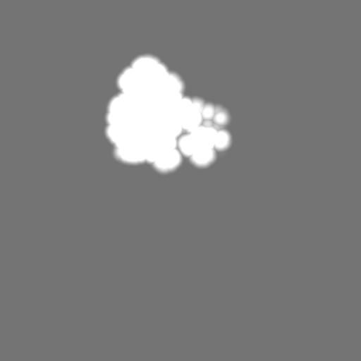
 Map data
 Sample
 Keywords
 Function and homology information
 Authors
 United States,
  Czech Republic, 4 items
  Citation


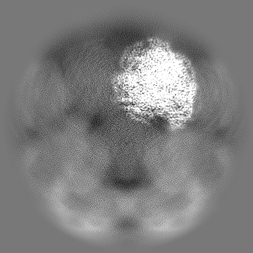
 Structure visualization
 Downloads & links
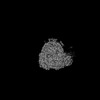
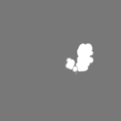
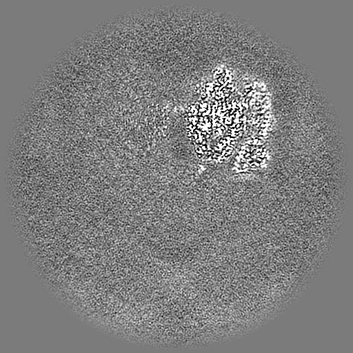
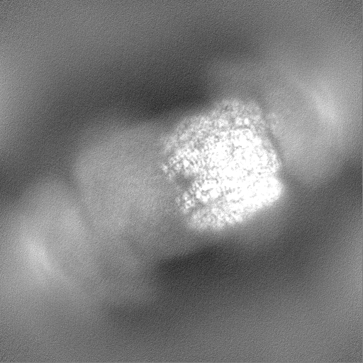
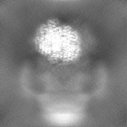
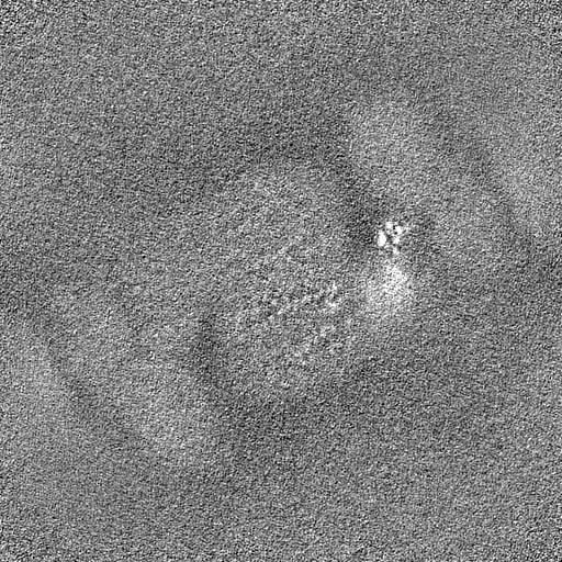
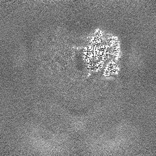
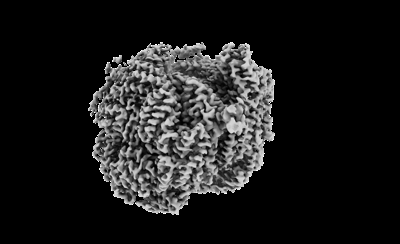 emd_41434.png
 http://ftp.pdbj.org/pub/emdb/structures/EMD-41434
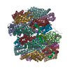


 Z (Sec.)
 Y (Row.)
 X (Col.)


 Sample components

 Processing
 Electron microscopy
 FIELD EMISSION GUN