Movie
Movie Controller


+ Open data

- Basic information
| Entry |  | |||||||||
|---|---|---|---|---|---|---|---|---|---|---|
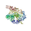
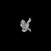
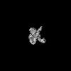
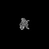
| Title | Tetrahymena Ribozyme scaffolded Zika Virus xrRNA | |||||||||
 Map data Map data | ||||||||||
 Sample Sample |
| |||||||||
 Keywords Keywords | Ribozyme / xrRNA / Scaffold / RNA | |||||||||
| Biological species |  Tetrahymena (eukaryote) / Tetrahymena (eukaryote) /   Zika virus Zika virus | |||||||||
| Method | single particle reconstruction / cryo EM / Resolution: 3.4 Å | |||||||||
 Authors Authors | Langeberg CJ / Kieft JS | |||||||||
| Funding support |  United States, 1 items United States, 1 items
| |||||||||
 Citation Citation | Journal: Nucleic Acids Res / Year: 2023 Title: A generalizable scaffold-based approach for structure determination of RNAs by cryo-EM. Authors: Conner J Langeberg / Jeffrey S Kieft / Abstract: Single-particle cryo-electron microscopy (cryo-EM) can reveal the structures of large and often dynamic molecules, but smaller biomolecules (≤50 kDa) remain challenging targets due to their ...Single-particle cryo-electron microscopy (cryo-EM) can reveal the structures of large and often dynamic molecules, but smaller biomolecules (≤50 kDa) remain challenging targets due to their intrinsic low signal to noise ratio. Methods to help resolve small proteins have been applied but development of similar approaches to aid in structural determination of small, structured RNA elements have lagged. Here, we present a scaffold-based approach that we used to recover maps of sub-25 kDa RNA domains to 4.5-5.0 Å. While lacking the detail of true high-resolution maps, these maps are suitable for model building and preliminary structure determination. We demonstrate this method helped faithfully recover the structure of several RNA elements of known structure, and that it promises to be generalized to other RNAs without disturbing their native fold. This approach may streamline the sample preparation process and reduce the optimization required for data collection. This first-generation scaffold approach provides a robust system to aid in RNA structure determination by cryo-EM and lays the groundwork for further scaffold optimization to achieve higher resolution. | |||||||||
| History |
|
- Structure visualization
| Supplemental images |
|---|

- Downloads & links
-EMDB archive
| Map data | emd_41308.map.gz | 255.6 MB |  EMDB map data format EMDB map data format | |
|---|---|---|---|---|
| Header (meta data) | emd-41308-v30.xmlemd-41308.xml | 16.8 KB 16.8 KB | Display Display | EMDB header |
| FSC (resolution estimation) | emd_41308_fsc.xml | 17 KB | Display | FSC data file |
| Images |  emd_41308.png emd_41308.png | 45 KB | ||
| Filedesc metadata | emd-41308.cif.gz | 5.7 KB | ||
| Others | emd_41308_half_map_1.map.gzemd_41308_half_map_2.map.gz | 474.9 MB 475 MB | ||
| Archive directory |  http://ftp.pdbj.org/pub/emdb/structures/EMD-41308ftp://ftp.pdbj.org/pub/emdb/structures/EMD-41308 http://ftp.pdbj.org/pub/emdb/structures/EMD-41308ftp://ftp.pdbj.org/pub/emdb/structures/EMD-41308 | HTTPS FTP |
-Related structure data
| Related structure data | 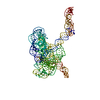 8tjqMC  8tjuC  8tjvC  8tjxC M: atomic model generated by this map C: citing same article ( |
|---|
-Links
| EMDB pages | EMDB (EBI/PDBe) / EMDataResource |
|---|
-Map
| File | Download / File: emd_41308.map.gz / Format: CCP4 / Size: 512 MB / Type: IMAGE STORED AS FLOATING POINT NUMBER (4 BYTES) | ||||||||||||||||||||||||||||||||||||
|---|---|---|---|---|---|---|---|---|---|---|---|---|---|---|---|---|---|---|---|---|---|---|---|---|---|---|---|---|---|---|---|---|---|---|---|---|---|
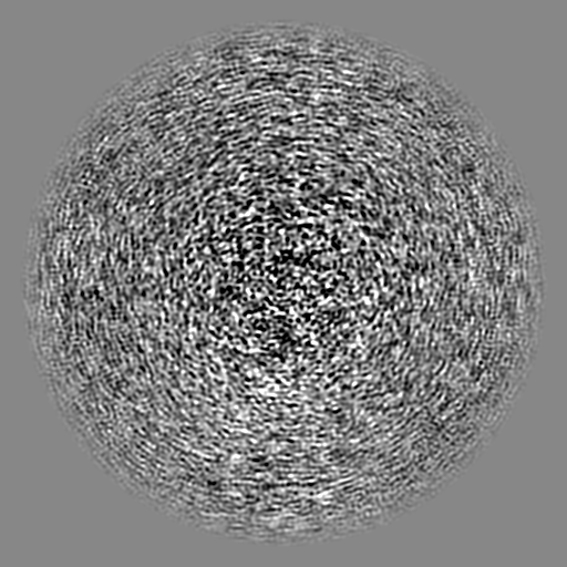
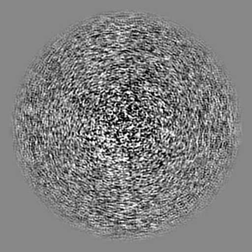
| Projections & slices | Image control
Images are generated by Spider. | ||||||||||||||||||||||||||||||||||||
| Voxel size | X=Y=Z: 0.8464 Å | ||||||||||||||||||||||||||||||||||||
| Density |
| ||||||||||||||||||||||||||||||||||||
| Symmetry | Space group: 1 | ||||||||||||||||||||||||||||||||||||
| Details | EMDB XML:
|
 Z (Sec.)
 Y (Row.)
 X (Col.)


-Supplemental data
-Half map: #2
| File | emd_41308_half_map_1.map | ||||||||||||
|---|---|---|---|---|---|---|---|---|---|---|---|---|---|
| Projections & Slices |
| ||||||||||||
| Density Histograms |


-Half map: #1
| File | emd_41308_half_map_2.map | ||||||||||||
|---|---|---|---|---|---|---|---|---|---|---|---|---|---|
| Projections & Slices |
| ||||||||||||
| Density Histograms |


- Sample components
-Entire : Tetrahymena Ribozyme scaffolded Zika Virus xrRNA
| Entire | Name: Tetrahymena Ribozyme scaffolded Zika Virus xrRNA |
|---|---|
| Components |
|
-Supramolecule #1: Tetrahymena Ribozyme scaffolded Zika Virus xrRNA
| Supramolecule | Name: Tetrahymena Ribozyme scaffolded Zika Virus xrRNA / type: complex / ID: 1 / Parent: 0 / Macromolecule list: #1 |
|---|---|
| Source (natural) | Organism: Tetrahymena (eukaryote) |
-Macromolecule #1: RNA (440-MER)
| Macromolecule | Name: RNA (440-MER) / type: rna / ID: 1 / Number of copies: 1 |
|---|---|
| Source (natural) | Organism: Zika virus |
| Molecular weight | Theoretical: 142.357188 KDa |
| Sequence | String: GGGUCAGGCC GGCUGAUAUG GAUGCAGUUC ACAGACUAAA UGUCGGUCGG GGAAGAUGUA UUCUUCUCAU AAGAUAUAGU CGGACCUCU CCUUAAUGGG AGCUAGCGGA UGAAGUGAUG CAACACUGGA GCCGCUGGGA ACUAAUUUGU AUGCGAAAGU A UAUUGAUU ...String: GGGUCAGGCC GGCUGAUAUG GAUGCAGUUC ACAGACUAAA UGUCGGUCGG GGAAGAUGUA UUCUUCUCAU AAGAUAUAGU CGGACCUCU CCUUAAUGGG AGCUAGCGGA UGAAGUGAUG CAACACUGGA GCCGCUGGGA ACUAAUUUGU AUGCGAAAGU A UAUUGAUU AGUUUUGGAG GAGGGAAAAG UUAUCAGGCA UGCACCUGGU AGCUAGUCUU UAAACCAAUA GAUUGCAUCG GU UUAAAAG GCAAGACCGU CAAAUUGCGG GAAAGGGGUC AACAGCCGUU CAGUACCAAG UCUCAGGGGA AACUUUGAGA UGG CCUUGC AAAGGGUAUG GUAAUAAGCU GACGGACAUG GUCCUAACCA CGCAGCCAAG UCCUAAGUCA GUCGCCACAG UUUG GGGAA AGCUGUGCAG CCUGUAACCC CCCCACGAAA GUGGG |
-Macromolecule #2: MAGNESIUM ION
| Macromolecule | Name: MAGNESIUM ION / type: ligand / ID: 2 / Number of copies: 28 / Formula: MG |
|---|---|
| Molecular weight | Theoretical: 24.305 Da |
-Experimental details
-Structure determination
| Method | cryo EM |
|---|---|
 Processing Processing | single particle reconstruction |
| Aggregation state | particle |
-Sample preparation
| Buffer | pH: 7.5 Component:
| |||||||||
|---|---|---|---|---|---|---|---|---|---|---|
| Grid | Model: C-flat-1.2/1.3 / Material: COPPER / Mesh: 50 / Pretreatment - Type: GLOW DISCHARGE / Pretreatment - Time: 6 sec. / Pretreatment - Atmosphere: OTHER | |||||||||
| Vitrification | Cryogen name: ETHANE |
- Electron microscopy
| Microscope | FEI TITAN KRIOS |
|---|---|
| Image recording | Film or detector model: GATAN K3 BIOQUANTUM (6k x 4k) / Average electron dose: 50.0 e/Å2 |
| Electron beam | Acceleration voltage: 300 kV / Electron source:  FIELD EMISSION GUN FIELD EMISSION GUN |
| Electron optics | Illumination mode: FLOOD BEAM / Imaging mode: BRIGHT FIELD / Nominal defocus max: 2.0 µm / Nominal defocus min: 0.8 µm |
| Experimental equipment |  Model: Titan Krios / Image courtesy: FEI Company |