 Movie
Movie Controller

[English] 日本語
 Yorodumi
- EMDB-37996: ParM present of genome of Desufitobacterium hafniense - Dc-cParM1 -
+ Open data

- Basic information
| Entry |  | ||||||||||||
|---|---|---|---|---|---|---|---|---|---|---|---|---|---|
| Title | ParM present of genome of Desufitobacterium hafniense - Dc-cParM1 | ||||||||||||
 Map data Map data | |||||||||||||
 Sample Sample |
| ||||||||||||
 Keywords Keywords | ParM / genome / segregation / plasmid / STRUCTURAL PROTEIN | ||||||||||||
| Function / homology | : / Archaeal actin homologue MreB-like, C-terminal / ParM-like / Actin-like protein, N-terminal / Actin like proteins N terminal domain / ATPase, nucleotide binding domain / Uncharacterized protein Function and homology information Function and homology information | ||||||||||||
| Biological species |  Desulfitobacterium hafniense Y51 (bacteria) Desulfitobacterium hafniense Y51 (bacteria) | ||||||||||||
| Method | helical reconstruction / cryo EM / Resolution: 4.0 Å | ||||||||||||
 Authors Authors | Ali S / Robinson RC / Narita A | ||||||||||||
| Funding support |  Japan, 3 items Japan, 3 items
| ||||||||||||
 Citation Citation | Journal: To Be Published Title: Bacterial genome encoded ParMs Authors: Ali S / Robinson RC / Narita A | ||||||||||||
| History |
|
- Structure visualization
| Supplemental images |
|---|

- Downloads & links
-EMDB archive
| Map data | emd_37996.map.gz | 116.4 MB | EMDB map data format | |
|---|---|---|---|---|
| Header (meta data) | emd-37996-v30.xmlemd-37996.xml | 18.7 KB 18.7 KB | Display Display | EMDB header |
| FSC (resolution estimation) | emd_37996_fsc.xml | 11.4 KB | Display | FSC data file |
| Images |  emd_37996.png emd_37996.png | 28.7 KB | ||
| Filedesc metadata | emd-37996.cif.gz | 6.2 KB | ||
| Others | emd_37996_additional_1.map.gzemd_37996_half_map_1.map.gzemd_37996_half_map_2.map.gz | 5.1 MB 97.8 MB 97.8 MB | ||
| Archive directory |  http://ftp.pdbj.org/pub/emdb/structures/EMD-37996ftp://ftp.pdbj.org/pub/emdb/structures/EMD-37996 http://ftp.pdbj.org/pub/emdb/structures/EMD-37996ftp://ftp.pdbj.org/pub/emdb/structures/EMD-37996 | HTTPS FTP |
-Validation report
| Summary document | emd_37996_validation.pdf.gz | 817.9 KB | Display | EMDB validaton report |
|---|---|---|---|---|
| Full document | emd_37996_full_validation.pdf.gz | 817.4 KB | Display | |
| Data in XML | emd_37996_validation.xml.gz | 18.7 KB | Display | |
| Data in CIF | emd_37996_validation.cif.gz | 24.8 KB | Display | |
| Arichive directory | https://ftp.pdbj.org/pub/emdb/validation_reports/EMD-37996ftp://ftp.pdbj.org/pub/emdb/validation_reports/EMD-37996 | HTTPS FTP |
-Related structure data
| Related structure data |  8x1iMC M: atomic model generated by this map C: citing same article ( |
|---|---|
| Similar structure data |
-Links
| EMDB pages | EMDB (EBI/PDBe) / EMDataResource |
|---|
-Map
| File | Download / File: emd_37996.map.gz / Format: CCP4 / Size: 125 MB / Type: IMAGE STORED AS FLOATING POINT NUMBER (4 BYTES) | ||||||||||||||||||||||||||||||||||||
|---|---|---|---|---|---|---|---|---|---|---|---|---|---|---|---|---|---|---|---|---|---|---|---|---|---|---|---|---|---|---|---|---|---|---|---|---|---|
| Projections & slices | Image control
Images are generated by Spider. | ||||||||||||||||||||||||||||||||||||
| Voxel size | X=Y=Z: 1.1 Å | ||||||||||||||||||||||||||||||||||||
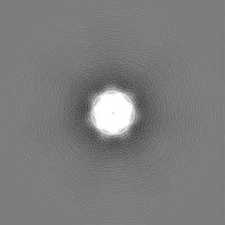
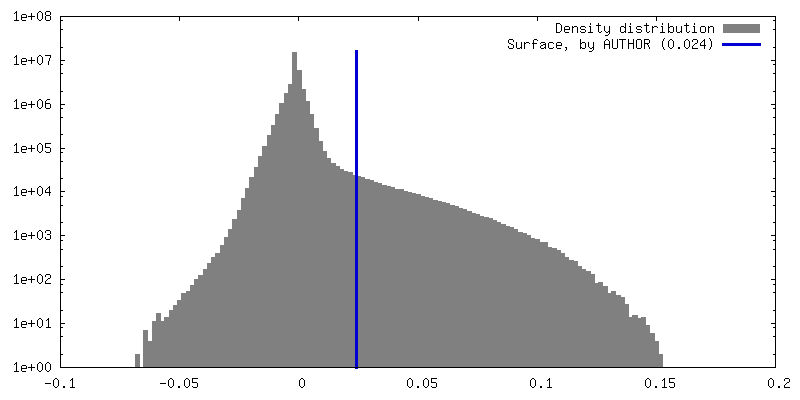
| Density |
| ||||||||||||||||||||||||||||||||||||
| Symmetry | Space group: 1 | ||||||||||||||||||||||||||||||||||||
| Details | EMDB XML:
|
 Z (Sec.)
 Y (Row.)
 X (Col.)


-Supplemental data
-Additional map: #1
| File | emd_37996_additional_1.map | ||||||||||||
|---|---|---|---|---|---|---|---|---|---|---|---|---|---|
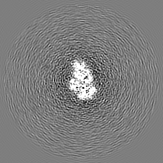
| Projections & Slices |
| ||||||||||||
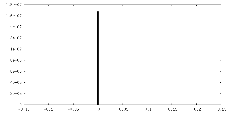
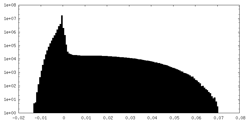
| Density Histograms |


-Half map: #2
| File | emd_37996_half_map_1.map | ||||||||||||
|---|---|---|---|---|---|---|---|---|---|---|---|---|---|
| Projections & Slices |
| ||||||||||||
| Density Histograms |


-Half map: #1
| File | emd_37996_half_map_2.map | ||||||||||||
|---|---|---|---|---|---|---|---|---|---|---|---|---|---|
| Projections & Slices |
| ||||||||||||
| Density Histograms |


- Sample components
-Entire : Dh-cParM1
| Entire | Name: Dh-cParM1 |
|---|---|
| Components |
|
-Supramolecule #1: Dh-cParM1
| Supramolecule | Name: Dh-cParM1 / type: organelle_or_cellular_component / ID: 1 / Parent: 0 / Macromolecule list: #1 Details: ParM present of genome of Desufitobacterium hafniense - Dh-cParM1 |
|---|---|
| Source (natural) | Organism: Desulfitobacterium hafniense Y51 (bacteria) |
| Molecular weight | Theoretical: 0.405 kDa/nm |
-Macromolecule #1: ParM present of genome of Desufitobacterium hafniense - Dh-cParM1
| Macromolecule | Name: ParM present of genome of Desufitobacterium hafniense - Dh-cParM1 type: protein_or_peptide / ID: 1 Details: genome encode ParM1 from Desulfitobacterium species Number of copies: 1 / Enantiomer: LEVO |
|---|---|
| Source (natural) | Organism: Desulfitobacterium hafniense Y51 (bacteria) |
| Molecular weight | Theoretical: 40.592141 KDa |
| Recombinant expression | Organism: |
| Sequence | String: MFENDILVAG GDPGFGAIKL DAGDTKVLFP AVICKGNERI FSALGNGNVS RGTDEEMQTG SLDVIVTNHS TGVSRHYFMG SLAESLNPN EAHYCWDEDK STDEEATALL VVALAVAQKE PKANIYLGTG VPVKYYAALK DKYEAELKGT WSVAFRSGPF K GQTRQLTI ...String: MFENDILVAG GDPGFGAIKL DAGDTKVLFP AVICKGNERI FSALGNGNVS RGTDEEMQTG SLDVIVTNHS TGVSRHYFMG SLAESLNPN EAHYCWDEDK STDEEATALL VVALAVAQKE PKANIYLGTG VPVKYYAALK DKYEAELKGT WSVAFRSGPF K GQTRQLTI IRSRVLPQSY GVFIKETLNE YGIPISPKLF NGYVVVIDPG FRTTDVATFY DGVMLDPPNS FSIEKGLKWA YT GVAEQLK EMTINHANPI ETDDKELDKV FRVNEGMYPW NNGAINLNPV MQDMLGQLGT DISREVKKSL KPMLGKIHTV LVA GKVGEM IFEHLQFENK VLIENPQFGN ATGFRIMAAN LVNNITKKAN AAP UniProtKB: Uncharacterized protein |
-Macromolecule #2: ADENOSINE-5'-DIPHOSPHATE
| Macromolecule | Name: ADENOSINE-5'-DIPHOSPHATE / type: ligand / ID: 2 / Number of copies: 1 / Formula: ADP |
|---|---|
| Molecular weight | Theoretical: 427.201 Da |
| Chemical component information |  ChemComp-ADP: |
-Macromolecule #3: MAGNESIUM ION
| Macromolecule | Name: MAGNESIUM ION / type: ligand / ID: 3 / Number of copies: 1 / Formula: MG |
|---|---|
| Molecular weight | Theoretical: 24.305 Da |
-Experimental details
-Structure determination
| Method | cryo EM |
|---|---|
 Processing Processing | helical reconstruction |
| Aggregation state | filament |
-Sample preparation
| Concentration | 10.0 mg/mL |
|---|---|
| Buffer | pH: 7.5 |
| Grid | Model: Quantifoil R1.2/1.3 / Material: MOLYBDENUM / Mesh: 200 / Support film - Material: CARBON / Pretreatment - Type: GLOW DISCHARGE / Pretreatment - Time: 40 sec. |
| Vitrification | Cryogen name: ETHANE / Chamber humidity: 90 % / Chamber temperature: 298 K / Instrument: LEICA EM GP |
| Details | ParM present of genome of Desufitobacterium hafniense - Dc-cParM1 |
- Electron microscopy
| Microscope | FEI TITAN KRIOS |
|---|---|
| Image recording | Film or detector model: FEI FALCON III (4k x 4k) / Detector mode: COUNTING / Average electron dose: 40.0 e/Å2 |
| Electron beam | Acceleration voltage: 300 kV / Electron source:  FIELD EMISSION GUN FIELD EMISSION GUN |
| Electron optics | Illumination mode: FLOOD BEAM / Imaging mode: BRIGHT FIELD / Nominal defocus max: 2.2 µm / Nominal defocus min: 1.5 µm |
| Experimental equipment |  Model: Titan Krios / Image courtesy: FEI Company |
+Image processing

-Atomic model buiding 1
| Initial model | Chain - Source name: AlphaFold / Chain - Initial model type: in silico model |
|---|---|
| Details | Initial fitting was performed using chimera and MDFF performed by ISOLDE and final real space refine by Phenix real space refinement. |
| Refinement | Space: REAL / Protocol: RIGID BODY FIT |
| Output model | PDB-8x1i: |
