Movie
Movie Controller

[English] 日本語
 Yorodumi
- EMDB-37752: Structure of the DDB1-AMBRA1 E3 ligase receptor complex linked to... -
+ Open data

- Basic information
| Entry |  | |||||||||
|---|---|---|---|---|---|---|---|---|---|---|
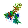
| Title | Structure of the DDB1-AMBRA1 E3 ligase receptor complex linked to cell cycle regulation | |||||||||
 Map data Map data | ||||||||||
 Sample Sample |
| |||||||||
 Keywords Keywords |  E3 ligase / STRUCTURAL PROTEIN E3 ligase / STRUCTURAL PROTEIN | |||||||||
| Function / homology |  Function and homology information Function and homology informationpositive regulation of free ubiquitin chain polymerization / : / response to mitochondrial depolarisation / positive regulation of mitophagy / autophagy of mitochondrion / positive regulation by virus of viral protein levels in host cell / epigenetic programming in the zygotic pronuclei / spindle assembly involved in female meiosis / positive regulation of regulatory T cell differentiation / Cul4-RING E3 ubiquitin ligase complex ...positive regulation of free ubiquitin chain polymerization / : / response to mitochondrial depolarisation / positive regulation of mitophagy / autophagy of mitochondrion / positive regulation by virus of viral protein levels in host cell / epigenetic programming in the zygotic pronuclei / spindle assembly involved in female meiosis / positive regulation of regulatory T cell differentiation / Cul4-RING E3 ubiquitin ligase complex / UV-damage excision repair / biological process involved in interaction with symbiont / neural tube development / negative regulation of cardiac muscle cell apoptotic process / regulation of mitotic cell cycle phase transition / WD40-repeat domain binding / Cul4A-RING E3 ubiquitin ligase complex / Cul4B-RING E3 ubiquitin ligase complex / ubiquitin ligase complex scaffold activity / Macroautophagy / negative regulation of reproductive process / negative regulation of developmental process / axoneme / regulation of G1/S transition of mitotic cell cycle / cullin family protein binding / mitophagy / autophagosome assembly / ubiquitin-like ligase-substrate adaptor activity / autophagosome / viral release from host cell / ectopic germ cell programmed cell death / positive regulation of viral genome replication / phagocytic vesicle / positive regulation of autophagy / positive regulation of protein dephosphorylation / positive regulation of gluconeogenesis / cellular response to starvation / proteasomal protein catabolic process / nucleotide-excision repair / Recognition of DNA damage by PCNA-containing replication complex / DNA Damage Recognition in GG-NER / regulation of circadian rhythm / Dual Incision in GG-NER / Transcription-Coupled Nucleotide Excision Repair (TC-NER) / Formation of TC-NER Pre-Incision Complex / Wnt signaling pathway / autophagy / Formation of Incision Complex in GG-NER / protein polyubiquitination / Dual incision in TC-NER / Gap-filling DNA repair synthesis and ligation in TC-NER / positive regulation of protein catabolic process / cellular response to UV / rhythmic process / protein-macromolecule adaptor activity / GTPase binding / site of double-strand break / Neddylation / ubiquitin-dependent protein catabolic process / proteasome-mediated ubiquitin-dependent protein catabolic process / protein phosphatase binding / negative regulation of neuron apoptotic process / mitochondrial outer membrane / chromosome, telomeric region / damaged DNA binding / cell differentiation / protein ubiquitination / cytoskeleton / cell cycle / negative regulation of cell population proliferation / focal adhesion / intracellular membrane-bounded organelle / DNA repair / apoptotic process / DNA damage response / ubiquitin protein ligase binding / protein-containing complex binding / nucleolus / regulation of transcription by RNA polymerase II / negative regulation of apoptotic process / perinuclear region of cytoplasm / endoplasmic reticulum / protein-containing complex / mitochondrion / DNA binding / extracellular space / extracellular exosome / nucleoplasm / nucleus / cytosol / cytoplasmSimilarity search - Function | |||||||||
| Biological species |  Homo sapiens (human) Homo sapiens (human) | |||||||||
| Method | single particle reconstruction / cryo EM / Resolution: 3.08 Å | |||||||||
 Authors Authors | Liu M / Wang Y / Su MY / Stjepanovic G | |||||||||
| Funding support |  China, 1 items China, 1 items
| |||||||||
 Citation Citation | Journal: Nat Commun / Year: 2023 Title: Structure of the DDB1-AMBRA1 E3 ligase receptor complex linked to cell cycle regulation. Authors: Ming Liu / Yang Wang / Fei Teng / Xinyi Mai / Xi Wang / Ming-Yuan Su / Goran Stjepanovic / Abstract: AMBRA1 is a tumor suppressor protein that functions as a substrate receptor of the ubiquitin conjugation system with roles in autophagy and the cell cycle regulatory network. The intrinsic disorder ...AMBRA1 is a tumor suppressor protein that functions as a substrate receptor of the ubiquitin conjugation system with roles in autophagy and the cell cycle regulatory network. The intrinsic disorder of AMBRA1 has thus far precluded its structural determination. To solve this problem, we analyzed the dynamics of AMBRA1 using hydrogen deuterium exchange mass spectrometry (HDX-MS). The HDX results indicated that AMBRA1 is a highly flexible protein and can be stabilized upon interaction with DDB1, the adaptor of the Cullin4A/B E3 ligase. Here, we present the cryo-EM structure of AMBRA1 in complex with DDB1 at 3.08 Å resolution. The structure shows that parts of the N- and C-terminal structural regions in AMBRA1 fold together into the highly dynamic WD40 domain and reveals how DDB1 engages with AMBRA1 to create a binding scaffold for substrate recruitment. The N-terminal helix-loop-helix motif and WD40 domain of AMBRA1 associate with the double-propeller fold of DDB1. We also demonstrate that DDB1 binding-defective AMBRA1 mutants prevent ubiquitination of the substrate Cyclin D1 in vitro and increase cell cycle progression. Together, these results provide structural insights into the AMBRA1-ubiquitin ligase complex and suggest a mechanism by which AMBRA1 acts as a hub involved in various physiological processes. | |||||||||
| History |
|
- Structure visualization
| Supplemental images |
|---|

- Downloads & links
-EMDB archive
| Map data | emd_37752.map.gz | 50.8 MB | EMDB map data format | |
|---|---|---|---|---|
| Header (meta data) | emd-37752-v30.xmlemd-37752.xml | 15.5 KB 15.5 KB | Display Display | EMDB header |
| Images | 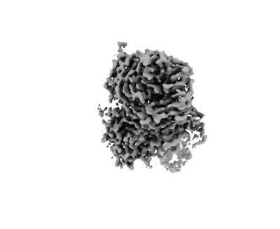 emd_37752.png emd_37752.png | 38 KB | ||
| Filedesc metadata | emd-37752.cif.gz | 6.1 KB | ||
| Others | emd_37752_half_map_1.map.gzemd_37752_half_map_2.map.gz | 95.5 MB 95.5 MB | ||
| Archive directory |  http://ftp.pdbj.org/pub/emdb/structures/EMD-37752ftp://ftp.pdbj.org/pub/emdb/structures/EMD-37752 http://ftp.pdbj.org/pub/emdb/structures/EMD-37752ftp://ftp.pdbj.org/pub/emdb/structures/EMD-37752 | HTTPS FTP |
-Related structure data
| Related structure data |  8wqrMC M: atomic model generated by this map C: citing same article ( |
|---|---|
| Similar structure data |
-Links
| EMDB pages | EMDB (EBI/PDBe) / EMDataResource |
|---|---|
| Related items in Molecule of the Month |


-Map
| File | Download / File: emd_37752.map.gz / Format: CCP4 / Size: 103 MB / Type: IMAGE STORED AS FLOATING POINT NUMBER (4 BYTES) | ||||||||||||||||||||
|---|---|---|---|---|---|---|---|---|---|---|---|---|---|---|---|---|---|---|---|---|---|
| Voxel size | X=Y=Z: 0.85 Å | ||||||||||||||||||||
| Density |
| ||||||||||||||||||||
| Symmetry | Space group: 1 | ||||||||||||||||||||
| Details | EMDB XML:
|
-Supplemental data
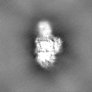
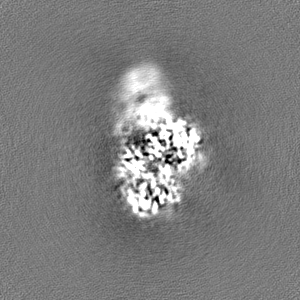
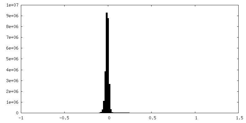
-Half map: #2
| File | emd_37752_half_map_1.map | ||||||||||||
|---|---|---|---|---|---|---|---|---|---|---|---|---|---|
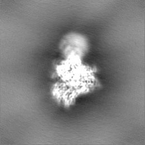
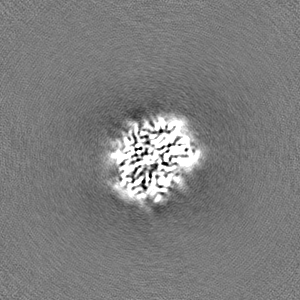
| Projections & Slices |
| ||||||||||||
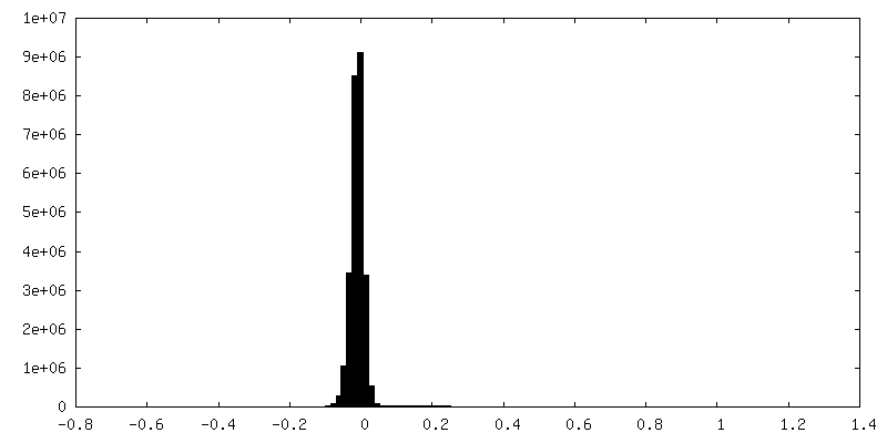
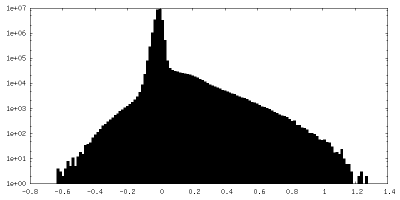
| Density Histograms |
 Z
Z Y
Y X
X


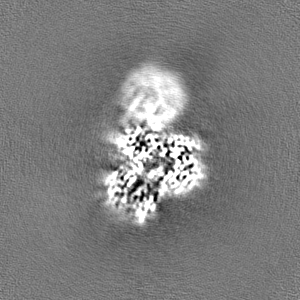
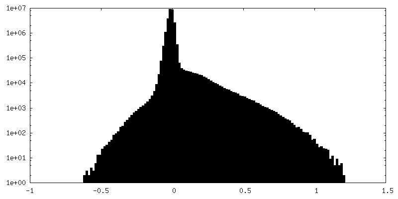
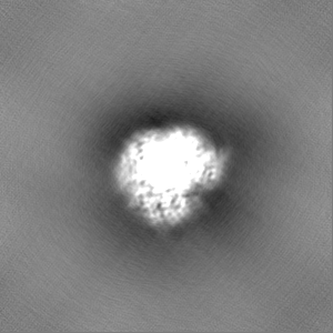
-Half map: #1
| File | emd_37752_half_map_2.map | ||||||||||||
|---|---|---|---|---|---|---|---|---|---|---|---|---|---|
| Projections & Slices |
| ||||||||||||
| Density Histograms |


- Sample components
-Entire : DDB1-AMBRA1 complex
| Entire | Name: DDB1-AMBRA1 complex |
|---|---|
| Components |
|
-Supramolecule #1: DDB1-AMBRA1 complex
| Supramolecule | Name: DDB1-AMBRA1 complex / type: complex / ID: 1 / Parent: 0 / Macromolecule list: all |
|---|---|
| Source (natural) | Organism: Homo sapiens (human) |
-Macromolecule #1: Activating molecule in BECN1-regulated autophagy protein 1
| Macromolecule | Name: Activating molecule in BECN1-regulated autophagy protein 1 type: protein_or_peptide / ID: 1 / Number of copies: 1 / Enantiomer: LEVO |
|---|---|
| Source (natural) | Organism: Homo sapiens (human) |
| Molecular weight | Theoretical: 44.496871 KDa |
| Recombinant expression | Organism: Homo sapiens (human) |
| Sequence | String: MKVVPEKNAV RILWGRERGA RAMGAQRLLQ ELVEDKTRWM KWEGKRVELP DSPRSTFLLA FSPDRTLLAS THVNHNIYIT EVKTGKCVH SLIGHRRTPW CVTFHPTISG LIASGCLDGE VRIWDLHGGS ESWFTDSNNA IASLAFHPTA QLLLIATANE I HFWDWSRR ...String: MKVVPEKNAV RILWGRERGA RAMGAQRLLQ ELVEDKTRWM KWEGKRVELP DSPRSTFLLA FSPDRTLLAS THVNHNIYIT EVKTGKCVH SLIGHRRTPW CVTFHPTISG LIASGCLDGE VRIWDLHGGS ESWFTDSNNA IASLAFHPTA QLLLIATANE I HFWDWSRR EPFAVVKTAS EMERVRLVRF DPLGHYLLTA IVNPSNSNIA NTTYRLQWWD FTKFDLPEIS NASVNVLVQN CK IYNDASC DISADGQLLA AFIPSSQRGF PDEGILAVYS LAPHNLGEML YTKRFGPNAI SVSLSPMGRY VMVGLASRRI LLH PSTEHM VAQVFRLQQA HGGETSMRRV FNVLYPMPAD QRRHVSINSA RWLPEPGLGL AYGTNKGDLV ICRPEALNSG UniProtKB: Activating molecule in BECN1-regulated autophagy protein 1, Activating molecule in BECN1-regulated autophagy protein 1 |
-Macromolecule #2: DNA damage-binding protein 1
| Macromolecule | Name: DNA damage-binding protein 1 / type: protein_or_peptide / ID: 2 / Number of copies: 1 / Enantiomer: LEVO |
|---|---|
| Source (natural) | Organism: Homo sapiens (human) |
| Molecular weight | Theoretical: 127.097469 KDa |
| Recombinant expression | Organism: Homo sapiens (human) |
| Sequence | String: MSYNYVVTAQ KPTAVNGCVT GHFTSAEDLN LLIAKNTRLE IYVVTAEGLR PVKEVGMYGK IAVMELFRPK GESKDLLFIL TAKYNACIL EYKQSGESID IITRAHGNVQ DRIGRPSETG IIGIIDPECR MIGLRLYDGL FKVIPLDRDN KELKAFNIRL E ELHVIDVK ...String: MSYNYVVTAQ KPTAVNGCVT GHFTSAEDLN LLIAKNTRLE IYVVTAEGLR PVKEVGMYGK IAVMELFRPK GESKDLLFIL TAKYNACIL EYKQSGESID IITRAHGNVQ DRIGRPSETG IIGIIDPECR MIGLRLYDGL FKVIPLDRDN KELKAFNIRL E ELHVIDVK FLYGCQAPTI CFVYQDPQGR HVKTYEVSLR EKEFNKGPWK QENVEAEASM VIAVPEPFGG AIIIGQESIT YH NGDKYLA IAPPIIKQST IVCHNRVDPN GSRYLLGDME GRLFMLLLEK EEQMDGTVTL KDLRVELLGE TSIAECLTYL DNG VVFVGS RLGDSQLVKL NVDSNEQGSY VVAMETFTNL GPIVDMCVVD LERQGQGQLV TCSGAFKEGS LRIIRNGIGI HEHA SIDLP GIKGLWPLRS DPNRETDDTL VLSFVGQTRV LMLNGEEVEE TELMGFVDDQ QTFFCGNVAH QQLIQITSAS VRLVS QEPK ALVSEWKEPQ AKNISVASCN SSQVVVAVGR ALYYLQIHPQ ELRQISHTEM EHEVACLDIT PLGDSNGLSP LCAIGL WTD ISARILKLPS FELLHKEMLG GEIIPRSILM TTFESSHYLL CALGDGALFY FGLNIETGLL SDRKKVTLGT QPTVLRT FR SLSTTNVFAC SDRPTVIYSS NHKLVFSNVN LKEVNYMCPL NSDGYPDSLA LANNSTLTIG TIDEIQKLHI RTVPLYES P RKICYQEVSQ CFGVLSSRIE VQDTSGGTTA LRPSASTQAL SSSVSSSKLF SSSTAPHETS FGEEVEVHNL LIIDQHTFE VLHAHQFLQN EYALSLVSCK LGKDPNTYFI VGTAMVYPEE AEPKQGRIVV FQYSDGKLQT VAEKEVKGAV YSMVEFNGKL LASINSTVR LYEWTTEKEL RTECNHYNNI MALYLKTKGD FILVGDLMRS VLLLAYKPME GNFEEIARDF NPNWMSAVEI L DDDNFLGA ENAFNLFVCQ KDSAATTDEE RQHLQEVGLF HLGEFVNVFC HGSLVMQNLG ETSTPTQGSV LFGTVNGMIG LV TSLSESW YNLLLDMQNR LNKVIKSVGK IEHSFWRSFH TERKTEPATG FIDGDLIESF LDISRPKMQE VVANLQYDDG SGM KREATA DDLIKVVEEL TRIH UniProtKB: DNA damage-binding protein 1 |
-Experimental details
-Structure determination
| Method | cryo EM |
|---|---|
 Processing Processing | single particle reconstruction |
| Aggregation state | particle |
-Sample preparation
| Buffer | pH: 7.4 |
|---|---|
| Vitrification | Cryogen name: ETHANE |
- Electron microscopy
| Microscope | FEI TITAN KRIOS |
|---|---|
| Electron beam | Acceleration voltage: 300 kV / Electron source: FIELD EMISSION GUN |
| Electron optics | Illumination mode: SPOT SCAN / Imaging mode: BRIGHT FIELDBright-field microscopy / Nominal defocus max: 1.8 µm / Nominal defocus min: 1.0 µm |
| Image recording | Film or detector model: GATAN K3 (6k x 4k) / Average electron dose: 58.96 e/Å2 |
| Experimental equipment |  Model: Titan Krios / Image courtesy: FEI Company |
-Image processing
| Startup model | Type of model: PDB ENTRY PDB model - PDB ID: |
|---|---|
| Initial angle assignment | Type: OTHER |
| Final angle assignment | Type: OTHER |
| Final reconstruction | Resolution.type: BY AUTHOR / Resolution: 3.08 Å / Resolution method: FSC 0.143 CUT-OFF / Number images used: 780090 |