+ Open data
- Basic information
| Entry |  | ||||||||||||
|---|---|---|---|---|---|---|---|---|---|---|---|---|---|
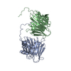
| Title | Cryo-EM structure of T. pseudonana PyShell helical tube | ||||||||||||
 Map data Map data | |||||||||||||
 Sample Sample |
| ||||||||||||
 Keywords Keywords | marine diatoms / photosynthesis / CO2-concentrating mechanism / pyrenoids / PLANT PROTEIN | ||||||||||||
| Function / homology | Uncharacterized protein Function and homology information Function and homology information | ||||||||||||
| Biological species |  Thalassiosira pseudonana (Diatom) Thalassiosira pseudonana (Diatom) | ||||||||||||
| Method | helical reconstruction / cryo EM / Resolution: 2.4 Å | ||||||||||||
 Authors Authors | Kawamoto A / Tohda R / Gerle C / Kurisu G | ||||||||||||
| Funding support |  Japan, 3 items Japan, 3 items
| ||||||||||||
 Citation Citation |  Journal: Cell / Year: 2024 Journal: Cell / Year: 2024Title: Diatom pyrenoids are encased in a protein shell that enables efficient CO fixation. Authors: Ginga Shimakawa / Manon Demulder / Serena Flori / Akihiro Kawamoto / Yoshinori Tsuji / Hermanus Nawaly / Atsuko Tanaka / Rei Tohda / Tadayoshi Ota / Hiroaki Matsui / Natsumi Morishima / ...Authors: Ginga Shimakawa / Manon Demulder / Serena Flori / Akihiro Kawamoto / Yoshinori Tsuji / Hermanus Nawaly / Atsuko Tanaka / Rei Tohda / Tadayoshi Ota / Hiroaki Matsui / Natsumi Morishima / Ryosuke Okubo / Wojciech Wietrzynski / Lorenz Lamm / Ricardo D Righetto / Clarisse Uwizeye / Benoit Gallet / Pierre-Henri Jouneau / Christoph Gerle / Genji Kurisu / Giovanni Finazzi / Benjamin D Engel / Yusuke Matsuda /     Abstract: Pyrenoids are subcompartments of algal chloroplasts that increase the efficiency of Rubisco-driven CO fixation. Diatoms fix up to 20% of global CO, but their pyrenoids remain poorly characterized. ...Pyrenoids are subcompartments of algal chloroplasts that increase the efficiency of Rubisco-driven CO fixation. Diatoms fix up to 20% of global CO, but their pyrenoids remain poorly characterized. Here, we used in vivo photo-crosslinking to identify pyrenoid shell (PyShell) proteins, which we localized to the pyrenoid periphery of model pennate and centric diatoms, Phaeodactylum tricornutum and Thalassiosira pseudonana. In situ cryo-electron tomography revealed that pyrenoids of both diatom species are encased in a lattice-like protein sheath. Single-particle cryo-EM yielded a 2.4-Å-resolution structure of an in vitro TpPyShell1 lattice, which showed how protein subunits interlock. T. pseudonana TpPyShell1/2 knockout mutants had no PyShell sheath, altered pyrenoid morphology, and a high-CO requiring phenotype, with reduced photosynthetic efficiency and impaired growth under standard atmospheric conditions. The structure and function of the diatom PyShell provide a molecular view of how CO is assimilated in the ocean, a critical ecosystem undergoing rapid change. | ||||||||||||
| History |
|
- Structure visualization
| Supplemental images |
|---|
- Downloads & links
-EMDB archive
| Map data |  emd_37751.map.gz emd_37751.map.gz | 1.2 MB |  EMDB map data format EMDB map data format | |
|---|---|---|---|---|
| Header (meta data) |  emd-37751-v30.xml emd-37751-v30.xml emd-37751.xml emd-37751.xml | 22.9 KB 22.9 KB | Display Display |  EMDB header EMDB header |
| FSC (resolution estimation) |  emd_37751_fsc.xml emd_37751_fsc.xml | 19 KB | Display |  FSC data file FSC data file |
| Images |  emd_37751.png emd_37751.png | 137.7 KB | ||
| Masks |  emd_37751_msk_1.map emd_37751_msk_1.map | 614.1 MB |  Mask map Mask map | |
| Filedesc metadata |  emd-37751.cif.gz emd-37751.cif.gz | 6.4 KB | ||
| Others |  emd_37751_additional_1.map.gz emd_37751_additional_1.map.gz emd_37751_additional_2.map.gz emd_37751_additional_2.map.gz emd_37751_half_map_1.map.gz emd_37751_half_map_1.map.gz emd_37751_half_map_2.map.gz emd_37751_half_map_2.map.gz | 61.8 MB 487.4 MB 491.4 MB 491.5 MB | ||
| Archive directory |  http://ftp.pdbj.org/pub/emdb/structures/EMD-37751 http://ftp.pdbj.org/pub/emdb/structures/EMD-37751 ftp://ftp.pdbj.org/pub/emdb/structures/EMD-37751 ftp://ftp.pdbj.org/pub/emdb/structures/EMD-37751 | HTTPS FTP |
-Validation report
| Summary document |  emd_37751_validation.pdf.gz emd_37751_validation.pdf.gz | 713.9 KB | Display |  EMDB validaton report EMDB validaton report |
|---|---|---|---|---|
| Full document |  emd_37751_full_validation.pdf.gz emd_37751_full_validation.pdf.gz | 713.4 KB | Display | |
| Data in XML |  emd_37751_validation.xml.gz emd_37751_validation.xml.gz | 26.6 KB | Display | |
| Data in CIF |  emd_37751_validation.cif.gz emd_37751_validation.cif.gz | 36 KB | Display | |
| Arichive directory |  https://ftp.pdbj.org/pub/emdb/validation_reports/EMD-37751 https://ftp.pdbj.org/pub/emdb/validation_reports/EMD-37751 ftp://ftp.pdbj.org/pub/emdb/validation_reports/EMD-37751 ftp://ftp.pdbj.org/pub/emdb/validation_reports/EMD-37751 | HTTPS FTP |
-Related structure data
| Related structure data |  8wqpMC M: atomic model generated by this map C: citing same article ( |
|---|---|
| Similar structure data | Similarity search - Function & homology  F&H Search F&H Search |
- Links
Links
| EMDB pages |  EMDB (EBI/PDBe) / EMDB (EBI/PDBe) /  EMDataResource EMDataResource |
|---|
- Map
Map
| File |  Download / File: emd_37751.map.gz / Format: CCP4 / Size: 614.1 MB / Type: IMAGE STORED AS FLOATING POINT NUMBER (4 BYTES) Download / File: emd_37751.map.gz / Format: CCP4 / Size: 614.1 MB / Type: IMAGE STORED AS FLOATING POINT NUMBER (4 BYTES) | ||||||||||||||||||||||||||||||||||||
|---|---|---|---|---|---|---|---|---|---|---|---|---|---|---|---|---|---|---|---|---|---|---|---|---|---|---|---|---|---|---|---|---|---|---|---|---|---|
| Projections & slices | Image control
Images are generated by Spider. | ||||||||||||||||||||||||||||||||||||
| Voxel size | X=Y=Z: 1.0875 Å | ||||||||||||||||||||||||||||||||||||
| Density |
| ||||||||||||||||||||||||||||||||||||
| Symmetry | Space group: 1 | ||||||||||||||||||||||||||||||||||||
| Details | EMDB XML:
|
-Supplemental data
-Mask #1
| File |  emd_37751_msk_1.map emd_37751_msk_1.map | ||||||||||||
|---|---|---|---|---|---|---|---|---|---|---|---|---|---|
| Projections & Slices |
| ||||||||||||
| Density Histograms |
-Additional map: #1
| File | emd_37751_additional_1.map | ||||||||||||
|---|---|---|---|---|---|---|---|---|---|---|---|---|---|
| Projections & Slices |
| ||||||||||||
| Density Histograms |
-Additional map: #2
| File | emd_37751_additional_2.map | ||||||||||||
|---|---|---|---|---|---|---|---|---|---|---|---|---|---|
| Projections & Slices |
| ||||||||||||
| Density Histograms |
-Half map: #1
| File | emd_37751_half_map_1.map | ||||||||||||
|---|---|---|---|---|---|---|---|---|---|---|---|---|---|
| Projections & Slices |
| ||||||||||||
| Density Histograms |
-Half map: #2
| File | emd_37751_half_map_2.map | ||||||||||||
|---|---|---|---|---|---|---|---|---|---|---|---|---|---|
| Projections & Slices |
| ||||||||||||
| Density Histograms |
- Sample components
-Entire : in vitro tube structure of T. pseudonana PyShell
| Entire | Name: in vitro tube structure of T. pseudonana PyShell |
|---|---|
| Components |
|
-Supramolecule #1: in vitro tube structure of T. pseudonana PyShell
| Supramolecule | Name: in vitro tube structure of T. pseudonana PyShell / type: complex / ID: 1 / Parent: 0 / Macromolecule list: all |
|---|---|
| Source (natural) | Organism:  Thalassiosira pseudonana (Diatom) Thalassiosira pseudonana (Diatom) |
-Macromolecule #1: Diatom the pyrenoid shell protein
| Macromolecule | Name: Diatom the pyrenoid shell protein / type: protein_or_peptide / ID: 1 / Number of copies: 2 / Enantiomer: LEVO |
|---|---|
| Source (natural) | Organism:  Thalassiosira pseudonana (Diatom) Thalassiosira pseudonana (Diatom) |
| Molecular weight | Theoretical: 24.98602 KDa |
| Recombinant expression | Organism:  |
| Sequence | String: (UNK)(UNK)(UNK)(UNK)(UNK)(UNK)(UNK)WDN SSPVIVQGGS LRTWSFANPA IESVQVLLKT EGRPLDADVE LW QGPDNTP HKMRVYVEDG ALRTFNAVIG TPRGPNTVAI RNIGQLEFPL DAVVRPDRDD GLAAGIASVA TRSETIQGGA LRT YPFNPT ...String: (UNK)(UNK)(UNK)(UNK)(UNK)(UNK)(UNK)WDN SSPVIVQGGS LRTWSFANPA IESVQVLLKT EGRPLDADVE LW QGPDNTP HKMRVYVEDG ALRTFNAVIG TPRGPNTVAI RNIGQLEFPL DAVVRPDRDD GLAAGIASVA TRSETIQGGA LRT YPFNPT VDSVAIILKT DGRPLNARIE LLQGPNNNKQ VVELYTEDGL DRPFFAIVET PGSGNVVRVV NTAPVEFPLY ASVD AYRVG GGGDWADDGL MIGRAF UniProtKB: Uncharacterized protein |
-Experimental details
-Structure determination
| Method | cryo EM |
|---|---|
 Processing Processing | helical reconstruction |
| Aggregation state | filament |
- Sample preparation
| Concentration | 2.0 mg/mL | |||||||||||||||
|---|---|---|---|---|---|---|---|---|---|---|---|---|---|---|---|---|
| Buffer | pH: 8 Component:
| |||||||||||||||
| Grid | Model: Quantifoil R1.2/1.3 / Material: COPPER / Mesh: 200 / Pretreatment - Type: GLOW DISCHARGE | |||||||||||||||
| Vitrification | Cryogen name: ETHANE / Chamber humidity: 100 % / Chamber temperature: 277 K / Instrument: FEI VITROBOT MARK IV |
- Electron microscopy
| Microscope | FEI TITAN KRIOS |
|---|---|
| Specialist optics | Spherical aberration corrector: Microscope was modified with a Cs corrector Energy filter - Name: GIF Bioquantum / Energy filter - Slit width: 20 eV |
| Image recording | Film or detector model: GATAN K3 (6k x 4k) / Number grids imaged: 1 / Number real images: 5951 / Average exposure time: 2.631 sec. / Average electron dose: 50.0 e/Å2 Details: Images were collected in movie-mode at 52 frames per second |
| Electron beam | Acceleration voltage: 300 kV / Electron source:  FIELD EMISSION GUN FIELD EMISSION GUN |
| Electron optics | C2 aperture diameter: 50.0 µm / Illumination mode: FLOOD BEAM / Imaging mode: BRIGHT FIELD / Cs: 0.01 mm / Nominal defocus max: 1.7 µm / Nominal defocus min: 0.5 µm / Nominal magnification: 81000 |
| Sample stage | Specimen holder model: FEI TITAN KRIOS AUTOGRID HOLDER / Cooling holder cryogen: NITROGEN |
| Experimental equipment |  Model: Titan Krios / Image courtesy: FEI Company |
+ Image processing
-Atomic model buiding 1
| Initial model | Chain - Source name: AlphaFold / Chain - Initial model type: in silico model |
|---|---|
| Refinement | Space: REAL / Protocol: AB INITIO MODEL |
| Output model |  PDB-8wqp: |
 Movie
Movie Controller


 Z (Sec.)
 Y (Row.)
 X (Col.)


