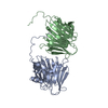
Journal: Cell / Year: 2024 Title: Diatom pyrenoids are encased in a protein shell that enables efficient CO fixation. Authors: Ginga Shimakawa / Manon Demulder / Serena Flori / Akihiro Kawamoto / Yoshinori Tsuji / Hermanus Nawaly / Atsuko Tanaka / Rei Tohda / Tadayoshi Ota / Hiroaki Matsui / Natsumi Morishima / ...Authors: Ginga Shimakawa / Manon Demulder / Serena Flori / Akihiro Kawamoto / Yoshinori Tsuji / Hermanus Nawaly / Atsuko Tanaka / Rei Tohda / Tadayoshi Ota / Hiroaki Matsui / Natsumi Morishima / Ryosuke Okubo / Wojciech Wietrzynski / Lorenz Lamm / Ricardo D Righetto / Clarisse Uwizeye / Benoit Gallet / Pierre-Henri Jouneau / Christoph Gerle / Genji Kurisu / Giovanni Finazzi / Benjamin D Engel / Yusuke Matsuda / Abstract: Pyrenoids are subcompartments of algal chloroplasts that increase the efficiency of Rubisco-driven CO fixation. Diatoms fix up to 20% of global CO, but their pyrenoids remain poorly characterized. ...Pyrenoids are subcompartments of algal chloroplasts that increase the efficiency of Rubisco-driven CO fixation. Diatoms fix up to 20% of global CO, but their pyrenoids remain poorly characterized. Here, we used in vivo photo-crosslinking to identify pyrenoid shell (PyShell) proteins, which we localized to the pyrenoid periphery of model pennate and centric diatoms, Phaeodactylum tricornutum and Thalassiosira pseudonana. In situ cryo-electron tomography revealed that pyrenoids of both diatom species are encased in a lattice-like protein sheath. Single-particle cryo-EM yielded a 2.4-Å-resolution structure of an in vitro TpPyShell1 lattice, which showed how protein subunits interlock. T. pseudonana TpPyShell1/2 knockout mutants had no PyShell sheath, altered pyrenoid morphology, and a high-CO requiring phenotype, with reduced photosynthetic efficiency and impaired growth under standard atmospheric conditions. The structure and function of the diatom PyShell provide a molecular view of how CO is assimilated in the ocean, a critical ecosystem undergoing rapid change.
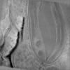
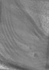
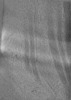
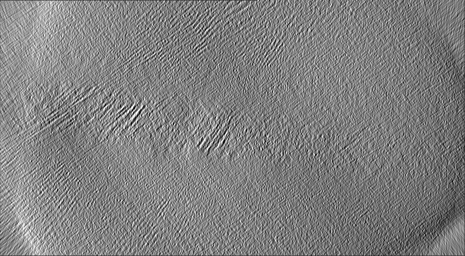
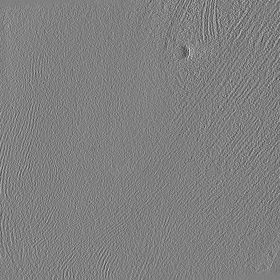
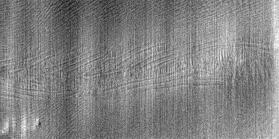
Name: T. pseudonana / type: cell / ID: 1 / Parent: 0 / Details: Pyrenoid in T. pseudonana chloroplast
Source (natural)
Organism: Thalassiosira pseudonana (Diatom)
-
Experimental details
-
Structure determination
Method
cryo EM
Processing
electron tomography
Aggregation state
cell
-
Sample preparation
Buffer
pH: 7
Vitrification
Cryogen name: ETHANE
Sectioning
Focused ion beam - Instrument: OTHER / Focused ion beam - Ion: OTHER / Focused ion beam - Voltage: 3 / Focused ion beam - Current: 0.03 / Focused ion beam - Duration: 600 / Focused ion beam - Temperature: 83 K / Focused ion beam - Initial thickness: 1000 / Focused ion beam - Final thickness: 150 Focused ion beam - Details: The value given for _em_focused_ion_beam.instrument is FEI Aquilos II. This is not in a list of allowed values {'OTHER', 'DB235'} so OTHER is written into the XML file.
-
Electron microscopy
Microscope
FEI TITAN KRIOS
Image recording
Film or detector model: FEI FALCON II (4k x 4k) / Detector mode: COUNTING / Average electron dose: 2.0 e/Å2
Electron beam
Acceleration voltage: 300 kV / Electron source: FIELD EMISSION GUN
In the structure databanks used in Yorodumi, some data are registered as the other names, "COVID-19 virus" and "2019-nCoV". Here are the details of the virus and the list of structure data.
Jan 31, 2019. EMDB accession codes are about to change! (news from PDBe EMDB page)
EMDB accession codes are about to change! (news from PDBe EMDB page)
The allocation of 4 digits for EMDB accession codes will soon come to an end. Whilst these codes will remain in use, new EMDB accession codes will include an additional digit and will expand incrementally as the available range of codes is exhausted. The current 4-digit format prefixed with “EMD-” (i.e. EMD-XXXX) will advance to a 5-digit format (i.e. EMD-XXXXX), and so on. It is currently estimated that the 4-digit codes will be depleted around Spring 2019, at which point the 5-digit format will come into force.
The EM Navigator/Yorodumi systems omit the EMD- prefix.
Related info.:Q: What is EMD? / ID/Accession-code notation in Yorodumi/EM Navigator
Yorodumi is a browser for structure data from EMDB, PDB, SASBDB, etc.
This page is also the successor to EM Navigator detail page, and also detail information page/front-end page for Omokage search.
The word "yorodu" (or yorozu) is an old Japanese word meaning "ten thousand". "mi" (miru) is to see.
Related info.:EMDB / PDB / SASBDB / Comparison of 3 databanks / Yorodumi Search / Aug 31, 2016. New EM Navigator & Yorodumi / Yorodumi Papers / Jmol/JSmol / Function and homology information / Changes in new EM Navigator and Yorodumi
 Movie
Movie Controller


 Open data
 Basic information
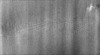
 Map data
 Sample
 Keywords
 Thalassiosira pseudonana (Diatom)
 Authors
 Citation


 Structure visualization
 Downloads & links
 EMDB map data format
 emd_18710.png
 http://ftp.pdbj.org/pub/emdb/structures/EMD-18710
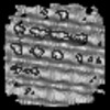
 Z (Sec.)
 Y (Row.)
 X (Col.)


 Sample components
 Processing
 Electron microscopy
 FIELD EMISSION GUN
