+ データを開く
- 基本情報
基本情報
| 登録情報 | データベース: EMDB / ID: EMD-3645 | |||||||||
|---|---|---|---|---|---|---|---|---|---|---|
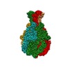
| タイトル | CryoEM density of TcdA1 in prepore state (SPHIRE tutorial) | |||||||||
 マップデータ マップデータ | CryoEM density of TcdA1 in prepore state | |||||||||
 試料 試料 |
| |||||||||
| 生物種 |  Photorhabdus luminescens (バクテリア) Photorhabdus luminescens (バクテリア) | |||||||||
| 手法 | 単粒子再構成法 / クライオ電子顕微鏡法 / 解像度: 3.5 Å | |||||||||
 データ登録者 データ登録者 | Raunser S / Gatsogiannis S / Roderer S | |||||||||
 引用 引用 |  ジャーナル: J Vis Exp / 年: 2017 ジャーナル: J Vis Exp / 年: 2017タイトル: High-resolution Single Particle Analysis from Electron Cryo-microscopy Images Using SPHIRE. 著者: Toshio Moriya / Michael Saur / Markus Stabrin / Felipe Merino / Horatiu Voicu / Zhong Huang / Pawel A Penczek / Stefan Raunser / Christos Gatsogiannis /   要旨: SPHIRE (SPARX for High-Resolution Electron Microscopy) is a novel open-source, user-friendly software suite for the semi-automated processing of single particle electron cryo-microscopy (cryo-EM) ...SPHIRE (SPARX for High-Resolution Electron Microscopy) is a novel open-source, user-friendly software suite for the semi-automated processing of single particle electron cryo-microscopy (cryo-EM) data. The protocol presented here describes in detail how to obtain a near-atomic resolution structure starting from cryo-EM micrograph movies by guiding users through all steps of the single particle structure determination pipeline. These steps are controlled from the new SPHIRE graphical user interface and require minimum user intervention. Using this protocol, a 3.5 Å structure of TcdA1, a Tc toxin complex from Photorhabdus luminescens, was derived from only 9500 single particles. This streamlined approach will help novice users without extensive processing experience and a priori structural information, to obtain noise-free and unbiased atomic models of their purified macromolecular complexes in their native state. | |||||||||
| 履歴 |
|
- 構造の表示
構造の表示
| ムービー |
 ムービービューア ムービービューア |
|---|---|
| 構造ビューア | EMマップ:  SurfView SurfView Molmil Molmil Jmol/JSmol Jmol/JSmol |
| 添付画像 |
- ダウンロードとリンク
-EMDBアーカイブ
| マップデータ |  emd_3645.map.gz emd_3645.map.gz | 6.4 MB |  EMDBマップデータ形式 EMDBマップデータ形式 | |
|---|---|---|---|---|
| ヘッダ (付随情報) |  emd-3645-v30.xml emd-3645-v30.xml emd-3645.xml emd-3645.xml | 11.5 KB 11.5 KB | 表示 表示 |  EMDBヘッダ EMDBヘッダ |
| 画像 |  emd_3645.png emd_3645.png | 90 KB | ||
| アーカイブディレクトリ |  http://ftp.pdbj.org/pub/emdb/structures/EMD-3645 http://ftp.pdbj.org/pub/emdb/structures/EMD-3645 ftp://ftp.pdbj.org/pub/emdb/structures/EMD-3645 ftp://ftp.pdbj.org/pub/emdb/structures/EMD-3645 | HTTPS FTP |
-検証レポート
| 文書・要旨 |  emd_3645_validation.pdf.gz emd_3645_validation.pdf.gz | 237.5 KB | 表示 |  EMDB検証レポート EMDB検証レポート |
|---|---|---|---|---|
| 文書・詳細版 |  emd_3645_full_validation.pdf.gz emd_3645_full_validation.pdf.gz | 236.7 KB | 表示 | |
| XML形式データ |  emd_3645_validation.xml.gz emd_3645_validation.xml.gz | 6.6 KB | 表示 | |
| アーカイブディレクトリ |  https://ftp.pdbj.org/pub/emdb/validation_reports/EMD-3645 https://ftp.pdbj.org/pub/emdb/validation_reports/EMD-3645 ftp://ftp.pdbj.org/pub/emdb/validation_reports/EMD-3645 ftp://ftp.pdbj.org/pub/emdb/validation_reports/EMD-3645 | HTTPS FTP |
-関連構造データ
| 関連構造データ | |
|---|---|
| 類似構造データ | |
| 電子顕微鏡画像生データ |  EMPIAR-10089 (タイトル: Single Particle cryoEM Dataset of TcdA1 in prepore state (SPHIRE tutorial) EMPIAR-10089 (タイトル: Single Particle cryoEM Dataset of TcdA1 in prepore state (SPHIRE tutorial)Data size: 72.8 Data #1: Unaligned multi-frame micrographs of TcdA1 in prepore state (SPHIRE tutorial dataset) [micrographs - multiframe]) |
- リンク
リンク
| EMDBのページ |  EMDB (EBI/PDBe) / EMDB (EBI/PDBe) /  EMDataResource EMDataResource |
|---|
- マップ
マップ
| ファイル |  ダウンロード / ファイル: emd_3645.map.gz / 形式: CCP4 / 大きさ: 166.4 MB / タイプ: IMAGE STORED AS FLOATING POINT NUMBER (4 BYTES) ダウンロード / ファイル: emd_3645.map.gz / 形式: CCP4 / 大きさ: 166.4 MB / タイプ: IMAGE STORED AS FLOATING POINT NUMBER (4 BYTES) | ||||||||||||||||||||||||||||||||||||||||||||||||||||||||||||
|---|---|---|---|---|---|---|---|---|---|---|---|---|---|---|---|---|---|---|---|---|---|---|---|---|---|---|---|---|---|---|---|---|---|---|---|---|---|---|---|---|---|---|---|---|---|---|---|---|---|---|---|---|---|---|---|---|---|---|---|---|---|
| 注釈 | CryoEM density of TcdA1 in prepore state | ||||||||||||||||||||||||||||||||||||||||||||||||||||||||||||
| 投影像・断面図 | 画像のコントロール
画像は Spider により作成 | ||||||||||||||||||||||||||||||||||||||||||||||||||||||||||||
| ボクセルのサイズ | X=Y=Z: 1.14 Å | ||||||||||||||||||||||||||||||||||||||||||||||||||||||||||||
| 密度 |
| ||||||||||||||||||||||||||||||||||||||||||||||||||||||||||||
| 対称性 | 空間群: 1 | ||||||||||||||||||||||||||||||||||||||||||||||||||||||||||||
| 詳細 | EMDB XML:
CCP4マップ ヘッダ情報:
| ||||||||||||||||||||||||||||||||||||||||||||||||||||||||||||
-添付データ
- 試料の構成要素
-全体 : TcdA1
| 全体 | 名称: TcdA1 |
|---|---|
| 要素 |
|
-超分子 #1: TcdA1
| 超分子 | 名称: TcdA1 / タイプ: complex / ID: 1 / 親要素: 0 / 含まれる分子: #1 / 詳細: Prepore state |
|---|---|
| 由来(天然) | 生物種:  Photorhabdus luminescens (バクテリア) Photorhabdus luminescens (バクテリア) |
| 組換発現 | 生物種:  |
| 分子量 | 理論値: 1.4 MDa |
-実験情報
-構造解析
| 手法 | クライオ電子顕微鏡法 |
|---|---|
 解析 解析 | 単粒子再構成法 |
| 試料の集合状態 | particle |
- 試料調製
試料調製
| 緩衝液 | pH: 5 構成要素:
| ||||||||
|---|---|---|---|---|---|---|---|---|---|
| グリッド | モデル: Quantifoil 2/1 2nm C / 材質: COPPER / メッシュ: 400 / 支持フィルム - 材質: CARBON / 支持フィルム - トポロジー: CONTINUOUS / 前処理 - タイプ: GLOW DISCHARGE | ||||||||
| 凍結 | 凍結剤: ETHANE / チャンバー内湿度: 90 % / 装置: GATAN CRYOPLUNGE 3 |
- 電子顕微鏡法
| 顕微鏡 | FEI TITAN KRIOS |
|---|---|
| 詳細 | Cs corrected microscope |
| 撮影 | フィルム・検出器のモデル: FEI FALCON II (4k x 4k) 実像数: 112 / 平均電子線量: 2.5 e/Å2 |
| 電子線 | 加速電圧: 300 kV / 電子線源:  FIELD EMISSION GUN FIELD EMISSION GUN |
| 電子光学系 | 照射モード: OTHER / 撮影モード: BRIGHT FIELD / 倍率(公称値): 59000 |
| 試料ステージ | ホルダー冷却材: NITROGEN |
| 実験機器 |  モデル: Titan Krios / 画像提供: FEI Company |
 ムービー
ムービー コントローラー


 Z (Sec.)
 Y (Row.)
 X (Col.)


