- EMDB-36119: Cryo-EM structure of viral topoisomerase in conformation 1 -
+
Open data
ID or keywords:
Loading...
-
Basic information
Entry
Database: EMDB / ID: EMD-36119
Title
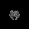
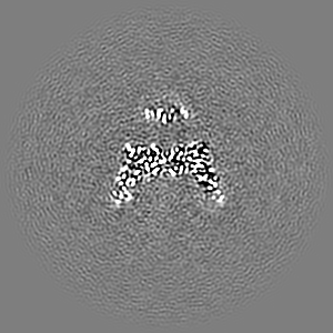
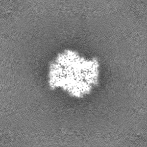
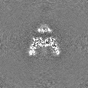
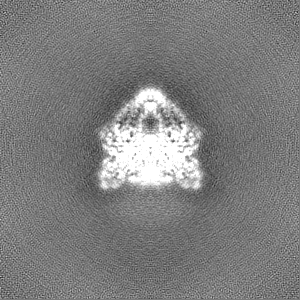
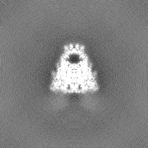
Cryo-EM structure of viral topoisomerase in conformation 1
Map data
Sample
Complex: P1192R
Protein or peptide: DNA topoisomerase 2
Keywords
topoisomerase / VIRAL PROTEIN
Function / homology
Function and homology information
sister chromatid segregation / DNA topoisomerase type II (double strand cut, ATP-hydrolyzing) activity / DNA topoisomerase (ATP-hydrolysing) / DNA topological change / host cell cytoplasm / DNA binding / ATP binding / metal ion binding Similarity search - Function
C-terminal associated domain of TOPRIM / C-terminal associated domain of TOPRIM / DNA topoisomerase II, eukaryotic-type / : / Topoisomerase (Topo) IIA-type catalytic domain profile. / DNA topoisomerase, type IIA, alpha-helical domain superfamily / DNA topoisomerase, type IIA, domain A / DNA topoisomerase, type IIA, domain A, alpha-beta / DNA gyrase/topoisomerase IV, subunit A / DNA Topoisomerase IV ...C-terminal associated domain of TOPRIM / C-terminal associated domain of TOPRIM / DNA topoisomerase II, eukaryotic-type / : / Topoisomerase (Topo) IIA-type catalytic domain profile. / DNA topoisomerase, type IIA, alpha-helical domain superfamily / DNA topoisomerase, type IIA, domain A / DNA topoisomerase, type IIA, domain A, alpha-beta / DNA gyrase/topoisomerase IV, subunit A / DNA Topoisomerase IV / DNA topoisomerase, type IIA / DNA topoisomerase, type IIA, conserved site / DNA topoisomerase II signature. / TopoisomeraseII / DNA topoisomerase, type IIA, subunit B, C-terminal / DNA topoisomerase, type IIA-like domain superfamily / Histidine kinase/HSP90-like ATPase superfamily / Ribosomal protein S5 domain 2-type fold, subgroup / Ribosomal protein S5 domain 2-type fold Similarity search - Domain/homology
Journal: mBio / Year: 2023 Title: Cryo-EM structures of African swine fever virus topoisomerase. Authors: Yan Zhao / Wenhua Kuang / Qiyin An / Jinyue Li / Yong Wang / Zengqin Deng / Abstract: African swine fever virus (ASFV) is a highly contagious virus that causes lethal hemorrhagic diseases known as African swine fever (ASF) with a case fatality rate of 100%. There is an urgent need to ...African swine fever virus (ASFV) is a highly contagious virus that causes lethal hemorrhagic diseases known as African swine fever (ASF) with a case fatality rate of 100%. There is an urgent need to develop anti-ASFV drugs. We determine the first high-resolution structures of viral topoisomerase ASFV P1192R in both the closed and open C-gate forms. P1192R shows a similar overall architecture with eukaryotic and prokaryotic type II topoisomerases, which have been successful targets of many antimicrobials and anticancer drugs, with the most similarity to yeast topo II. P1192R also exhibits differences in the details of active site configuration, which are important to enzyme activity. These two structures offer useful structural information for antiviral drug design and provide structural evidence to support that eukaryotic type IIA topoisomerase likely originated from horizontal gene transfer from the virus.
In the structure databanks used in Yorodumi, some data are registered as the other names, "COVID-19 virus" and "2019-nCoV". Here are the details of the virus and the list of structure data.
Jan 31, 2019. EMDB accession codes are about to change! (news from PDBe EMDB page)
EMDB accession codes are about to change! (news from PDBe EMDB page)
The allocation of 4 digits for EMDB accession codes will soon come to an end. Whilst these codes will remain in use, new EMDB accession codes will include an additional digit and will expand incrementally as the available range of codes is exhausted. The current 4-digit format prefixed with “EMD-” (i.e. EMD-XXXX) will advance to a 5-digit format (i.e. EMD-XXXXX), and so on. It is currently estimated that the 4-digit codes will be depleted around Spring 2019, at which point the 5-digit format will come into force.
The EM Navigator/Yorodumi systems omit the EMD- prefix.
Related info.:Q: What is EMD? / ID/Accession-code notation in Yorodumi/EM Navigator
Yorodumi is a browser for structure data from EMDB, PDB, SASBDB, etc.
This page is also the successor to EM Navigator detail page, and also detail information page/front-end page for Omokage search.
The word "yorodu" (or yorozu) is an old Japanese word meaning "ten thousand". "mi" (miru) is to see.
Related info.:EMDB / PDB / SASBDB / Comparison of 3 databanks / Yorodumi Search / Aug 31, 2016. New EM Navigator & Yorodumi / Yorodumi Papers / Jmol/JSmol / Function and homology information / Changes in new EM Navigator and Yorodumi
 Movie
Movie Controller


 Open data
 Basic information
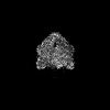
 Map data
 Sample
 Keywords
 Function and homology information
 African swine fever virus LIS57
 Authors
 Citation
 Structure visualization
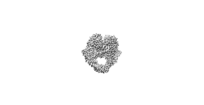
 Downloads & links
 emd_36119.png
 http://ftp.pdbj.org/pub/emdb/structures/EMD-36119


 Z (Sec.)
 Y (Row.)
 X (Col.)


 Sample components
 Komagataella pastoris (fungus)
 Processing
 Electron microscopy
 FIELD EMISSION GUN