Movie
Movie Controller


+ Open data

- Basic information
| Entry | 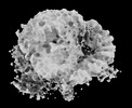 | |||||||||
|---|---|---|---|---|---|---|---|---|---|---|
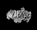
| Title | Concensus map of the Rpd3S-nucleosome complex | |||||||||
 Map data Map data | Concensus map | |||||||||
 Sample Sample |
| |||||||||
 Keywords Keywords | Histone deacetylase-nucleosome complex / GENE REGULATION | |||||||||
| Biological species |  | |||||||||
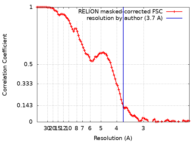
| Method | single particle reconstruction / cryo EM / Resolution: 3.7 Å | |||||||||
 Authors Authors | Cui H / Wang H | |||||||||
| Funding support |  China, 1 items China, 1 items
| |||||||||
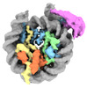
 Citation Citation | Journal: Nat Struct Mol Biol / Year: 2023 Title: Structure of histone deacetylase complex Rpd3S bound to nucleosome. Authors: Wulong Li / Hengjun Cui / Zhimin Lu / Haibo Wang / Abstract: Crosstalk between histone modifications represents a fundamental epigenetic mechanism in gene regulation. During the transcription elongation process, the histone deacetylase complex Rpd3S is ...Crosstalk between histone modifications represents a fundamental epigenetic mechanism in gene regulation. During the transcription elongation process, the histone deacetylase complex Rpd3S is recruited to H3K36-methylated nucleosomes to suppress cryptic transcription initiation. However, how subunits of Rpd3S are assembled and coordinated to recognize nucleosomal substrates and exert their deacetylation function remains unclear. Here we report the structure of Saccharomyces cerevisiae Rpd3S deacetylase bound to H3K36me3-modified nucleosome at 3.1 Å resolution. It shows that Sin3 and Rco1 subunits orchestrate the assembly of the complex and mediate its contact with nucleosome at multiple sites, with the Sin3-DNA interface as a pivotal anchor. The PHD1 domain of Rco1 recognizes the unmodified H3K4 and places the following H3 tail toward the active site of Rpd3, while the chromodomain of Eaf3 subunit recognizes the H3K36me3 mark and contacts both nucleosomal and linker DNA. The second copy of Eaf3-Rco1 is involved in neighboring nucleosome binding. Our work unravels the structural basis of chromatin targeting and deacetylation by the Rpd3S complex. | |||||||||
| History |
|
- Structure visualization
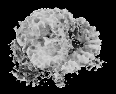
| Supplemental images |
|---|
- Downloads & links
-EMDB archive
| Map data | emd_35217.map.gz | 49.3 MB |  EMDB map data format EMDB map data format | |
|---|---|---|---|---|
| Header (meta data) | emd-35217-v30.xmlemd-35217.xml | 12.7 KB 12.7 KB | Display Display | EMDB header |
| FSC (resolution estimation) | emd_35217_fsc.xml | 9.1 KB | Display | FSC data file |
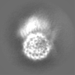
| Images |  emd_35217.png emd_35217.png | 73.3 KB | ||
| Filedesc metadata | emd-35217.cif.gz | 3.9 KB | ||
| Others | emd_35217_half_map_1.map.gzemd_35217_half_map_2.map.gz | 49.6 MB 49.6 MB | ||
| Archive directory |  http://ftp.pdbj.org/pub/emdb/structures/EMD-35217ftp://ftp.pdbj.org/pub/emdb/structures/EMD-35217 http://ftp.pdbj.org/pub/emdb/structures/EMD-35217ftp://ftp.pdbj.org/pub/emdb/structures/EMD-35217 | HTTPS FTP |
-Validation report
| Summary document | emd_35217_validation.pdf.gz | 715.1 KB | Display | EMDB validaton report |
|---|---|---|---|---|
| Full document | emd_35217_full_validation.pdf.gz | 714.7 KB | Display | |
| Data in XML | emd_35217_validation.xml.gz | 16.3 KB | Display | |
| Data in CIF | emd_35217_validation.cif.gz | 21.3 KB | Display | |
| Arichive directory | https://ftp.pdbj.org/pub/emdb/validation_reports/EMD-35217ftp://ftp.pdbj.org/pub/emdb/validation_reports/EMD-35217 | HTTPS FTP |
-Related structure data


-Links
| EMDB pages | EMDB (EBI/PDBe) / EMDataResource |
|---|
-Map
| File | Download / File: emd_35217.map.gz / Format: CCP4 / Size: 64 MB / Type: IMAGE STORED AS FLOATING POINT NUMBER (4 BYTES) | ||||||||||||||||||||
|---|---|---|---|---|---|---|---|---|---|---|---|---|---|---|---|---|---|---|---|---|---|
| Annotation | Concensus map | ||||||||||||||||||||
| Voxel size | X=Y=Z: 1.05 Å | ||||||||||||||||||||
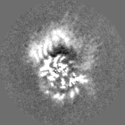
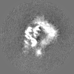
| Density |
| ||||||||||||||||||||
| Symmetry | Space group: 1 | ||||||||||||||||||||
| Details | EMDB XML:
|
-Supplemental data
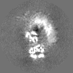
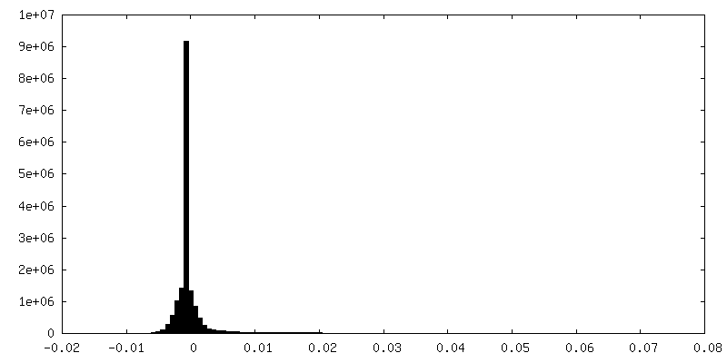
-Half map: EM half1 map
| File | emd_35217_half_map_1.map | ||||||||||||
|---|---|---|---|---|---|---|---|---|---|---|---|---|---|
| Annotation | EM half1 map | ||||||||||||
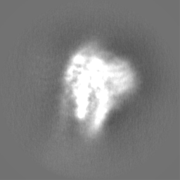
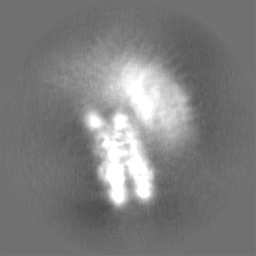
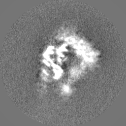
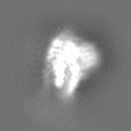
| Projections & Slices |
| ||||||||||||
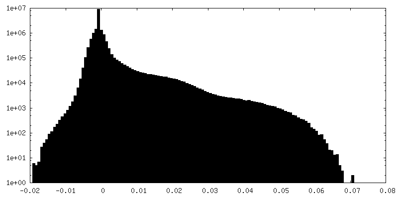
| Density Histograms |
 Z
Z Y
Y X
X


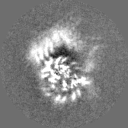
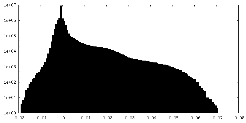
-Half map: EM half2 map
| File | emd_35217_half_map_2.map | ||||||||||||
|---|---|---|---|---|---|---|---|---|---|---|---|---|---|
| Annotation | EM half2 map | ||||||||||||
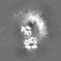
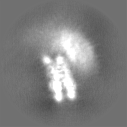
| Projections & Slices |
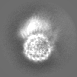
| ||||||||||||
| Density Histograms |
- Sample components
-Entire : Rpd3S histone deacetylase in complex with nucleosome
| Entire | Name: Rpd3S histone deacetylase in complex with nucleosome |
|---|---|
| Components |
|
-Supramolecule #1: Rpd3S histone deacetylase in complex with nucleosome
| Supramolecule | Name: Rpd3S histone deacetylase in complex with nucleosome / type: complex / ID: 1 / Parent: 0 |
|---|---|
| Source (natural) | Organism: |
-Experimental details
-Structure determination
| Method | cryo EM |
|---|---|
 Processing Processing | single particle reconstruction |
| Aggregation state | particle |
-Sample preparation
| Buffer | pH: 7.5 Details: 20 mM HEPES-Na pH 7.5, 40 mM KCl, 2 mM MgCl2, 1 mM TCEP |
|---|---|
| Vitrification | Cryogen name: ETHANE / Chamber humidity: 100 % / Chamber temperature: 277.15 K / Instrument: FEI VITROBOT MARK IV |
- Electron microscopy
| Microscope | FEI TITAN KRIOS |
|---|---|
| Specialist optics | Energy filter - Name: GIF Bioquantum / Energy filter - Slit width: 20 eV |
| Image recording | Film or detector model: GATAN K3 BIOQUANTUM (6k x 4k) / Average electron dose: 44.0 e/Å2 |
| Electron beam | Acceleration voltage: 300 kV / Electron source:  FIELD EMISSION GUN FIELD EMISSION GUN |
| Electron optics | Illumination mode: FLOOD BEAM / Imaging mode: BRIGHT FIELD / Nominal defocus max: 2.0 µm / Nominal defocus min: 0.8 µm |
| Sample stage | Specimen holder model: FEI TITAN KRIOS AUTOGRID HOLDER / Cooling holder cryogen: NITROGEN |
| Experimental equipment |  Model: Titan Krios / Image courtesy: FEI Company |