 Movie
Movie Controller


+ Open data

- Basic information
| Entry | Database: PDB / ID: 8hxx | ||||||
|---|---|---|---|---|---|---|---|
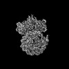
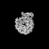
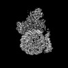
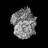
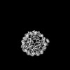
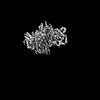
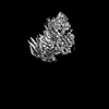
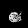
| Title | Cryo-EM structure of the histone deacetylase complex Rpd3S | ||||||
 Components Components |
| ||||||
 Keywords Keywords | GENE REGULATION / Histone deacetylase complex | ||||||
| Function / homology |  Function and homology information Function and homology informationSnt2C complex / negative regulation of silent mating-type cassette heterochromatin formation / negative regulation of reciprocal meiotic recombination / Rpd3L complex / protein localization to nucleolar rDNA repeats / negative regulation of rDNA heterochromatin formation / Rpd3L-Expanded complex / Rpd3S complex / rDNA chromatin condensation / nucleophagy ...Snt2C complex / negative regulation of silent mating-type cassette heterochromatin formation / negative regulation of reciprocal meiotic recombination / Rpd3L complex / protein localization to nucleolar rDNA repeats / negative regulation of rDNA heterochromatin formation / Rpd3L-Expanded complex / Rpd3S complex / rDNA chromatin condensation / nucleophagy / HDACs deacetylate histones / histone deacetylase activity, hydrolytic mechanism / histone deacetylase / SUMOylation of chromatin organization proteins / cellular response to nitrogen starvation / regulation of DNA-templated DNA replication initiation / negative regulation of transcription by RNA polymerase I / histone deacetylase activity / Sin3-type complex / NuA4 histone acetyltransferase complex / Estrogen-dependent gene expression / histone deacetylase complex / positive regulation of macroautophagy / nuclear periphery / meiotic cell cycle / transcription elongation by RNA polymerase II / G1/S transition of mitotic cell cycle / double-strand break repair via nonhomologous end joining / G2/M transition of mitotic cell cycle / structural constituent of chromatin / transcription corepressor activity / nucleosome / heterochromatin formation / nucleosome assembly / cellular response to heat / response to oxidative stress / transcription coactivator activity / chromatin remodeling / protein heterodimerization activity / cell division / DNA repair / regulation of DNA-templated transcription / regulation of transcription by RNA polymerase II / chromatin / negative regulation of transcription by RNA polymerase II / positive regulation of transcription by RNA polymerase II / DNA binding / metal ion binding / identical protein binding / nucleus / cytoplasm Similarity search - Function | ||||||
| Biological species |  | ||||||
| Method | ELECTRON MICROSCOPY / single particle reconstruction / cryo EM / Resolution: 3 Å | ||||||
 Authors Authors | Cui, H. / Wang, H. | ||||||
| Funding support |  China, 1items China, 1items
| ||||||
 Citation Citation | Journal: Nat Struct Mol Biol / Year: 2023 Title: Structure of histone deacetylase complex Rpd3S bound to nucleosome. Authors: Wulong Li / Hengjun Cui / Zhimin Lu / Haibo Wang / Abstract: Crosstalk between histone modifications represents a fundamental epigenetic mechanism in gene regulation. During the transcription elongation process, the histone deacetylase complex Rpd3S is ...Crosstalk between histone modifications represents a fundamental epigenetic mechanism in gene regulation. During the transcription elongation process, the histone deacetylase complex Rpd3S is recruited to H3K36-methylated nucleosomes to suppress cryptic transcription initiation. However, how subunits of Rpd3S are assembled and coordinated to recognize nucleosomal substrates and exert their deacetylation function remains unclear. Here we report the structure of Saccharomyces cerevisiae Rpd3S deacetylase bound to H3K36me3-modified nucleosome at 3.1 Å resolution. It shows that Sin3 and Rco1 subunits orchestrate the assembly of the complex and mediate its contact with nucleosome at multiple sites, with the Sin3-DNA interface as a pivotal anchor. The PHD1 domain of Rco1 recognizes the unmodified H3K4 and places the following H3 tail toward the active site of Rpd3, while the chromodomain of Eaf3 subunit recognizes the H3K36me3 mark and contacts both nucleosomal and linker DNA. The second copy of Eaf3-Rco1 is involved in neighboring nucleosome binding. Our work unravels the structural basis of chromatin targeting and deacetylation by the Rpd3S complex. | ||||||
| History |
|
- Structure visualization
| Structure viewer | Molecule: MolmilJmol/JSmol |
|---|
- Downloads & links
-Download
| PDBx/mmCIF format | 8hxx.cif.gz | 424.7 KB | Display | PDBx/mmCIF format |
|---|---|---|---|---|
| PDB format | pdb8hxx.ent.gz | 312.5 KB | Display | PDB format |
| PDBx/mmJSON format | 8hxx.json.gz | Tree view | PDBx/mmJSON format | |
| Others |  Other downloads Other downloads |
-Validation report
| Summary document | 8hxx_validation.pdf.gz | 427.9 KB | Display | wwPDB validaton report |
|---|---|---|---|---|
| Full document | 8hxx_full_validation.pdf.gz | 450.5 KB | Display | |
| Data in XML | 8hxx_validation.xml.gz | 36.9 KB | Display | |
| Data in CIF | 8hxx_validation.cif.gz | 55.9 KB | Display | |
| Arichive directory | https://data.pdbj.org/pub/pdb/validation_reports/hx/8hxxftp://data.pdbj.org/pub/pdb/validation_reports/hx/8hxx | HTTPS FTP |
-Related structure data
| Related structure data |  35081MC  8hxyC  8hxzC  8hy0C  8jhoC M: map data used to model this data C: citing same article ( |
|---|---|
| Similar structure data |
-Links
 PDBj
PDBj


- Assembly
| Deposited unit | 
|
|---|---|
| 1 |
|
-Components
-Protein , 5 types, 7 molecules KLMONPE
| #1: Protein | Mass: 175047.266 Da / Num. of mol.: 1 Source method: isolated from a genetically manipulated source Source: (gene. exp.) Gene: SIN3, CPE1, GAM2, RPD1, SDI1, SDS16, UME4, YOL004W / Production host:  Trichoplusia ni (cabbage looper) / References: UniProt: P22579 Trichoplusia ni (cabbage looper) / References: UniProt: P22579 | ||||
|---|---|---|---|---|---|
| #2: Protein | Mass: 48961.957 Da / Num. of mol.: 1 Source method: isolated from a genetically manipulated source Source: (gene. exp.) Gene: RPD3, MOF6, REC3, SDI2, SDS6, YNL330C, N0305 / Production host: Trichoplusia ni (cabbage looper) / References: UniProt: P32561, histone deacetylase | ||||
| #3: Protein | Mass: 45266.406 Da / Num. of mol.: 2 Source method: isolated from a genetically manipulated source Source: (gene. exp.) Gene: EAF3 / Production host: Trichoplusia ni (cabbage looper) / References: UniProt: A0A8H4F719#4: Protein | Mass: 78951.305 Da / Num. of mol.: 2 Source method: isolated from a genetically manipulated source Source: (gene. exp.) Gene: RCO1 / Production host: Trichoplusia ni (cabbage looper) / References: UniProt: A0A8H4BXB0#5: Protein | | Mass: 15331.982 Da / Num. of mol.: 1 / Mutation: C110A Source method: isolated from a genetically manipulated source Details: Author stated that Cys110 residue of chain E was mutated to Ala due to the preparation of ML3-modified nucleosome. Source: (gene. exp.)  |
-Non-polymers , 1 types, 7 molecules 
| #6: Chemical | ChemComp-ZN /  Mass: 65.409 Da / Num. of mol.: 7 / Source method: obtained synthetically / Formula: Zn / Feature type: SUBJECT OF INVESTIGATION Mass: 65.409 Da / Num. of mol.: 7 / Source method: obtained synthetically / Formula: Zn / Feature type: SUBJECT OF INVESTIGATION |
|---|
-Details
| Has ligand of interest | Y |
|---|---|
| Has protein modification | N |
-Experimental details
-Experiment
| Experiment | Method: ELECTRON MICROSCOPY |
|---|---|
| EM experiment | Aggregation state: PARTICLE / 3D reconstruction method: single particle reconstruction |
- Sample preparation
| Component | Name: Rpd3S histone deacetylase in complex with histone H3 tail Type: COMPLEX / Entity ID: #1-#5 / Source: MULTIPLE SOURCES |
|---|---|
| Molecular weight | Experimental value: NO |
| Source (natural) | Organism: |
| Buffer solution | pH: 7.5 Details: 20 mM HEPES-Na pH 7.5, 40 mM KCl, 2 mM MgCl2, 1 mM TCEP |
| Specimen | Embedding applied: NO / Shadowing applied: NO / Staining applied: NO / Vitrification applied: YES |
| Vitrification | Instrument: FEI VITROBOT MARK IV / Cryogen name: ETHANE / Humidity: 100 % / Chamber temperature: 277.15 K |
- Electron microscopy imaging
| Experimental equipment |  Model: Titan Krios / Image courtesy: FEI Company |
|---|---|
| Microscopy | Model: FEI TITAN KRIOS |
| Electron gun | Electron source:  FIELD EMISSION GUN / Accelerating voltage: 300 kV / Illumination mode: FLOOD BEAM FIELD EMISSION GUN / Accelerating voltage: 300 kV / Illumination mode: FLOOD BEAM |
| Electron lens | Mode: BRIGHT FIELD / Nominal defocus max: 2000 nm / Nominal defocus min: 800 nm |
| Specimen holder | Cryogen: NITROGEN / Specimen holder model: FEI TITAN KRIOS AUTOGRID HOLDER |
| Image recording | Electron dose: 44 e/Å2 / Film or detector model: GATAN K3 BIOQUANTUM (6k x 4k) |
| EM imaging optics | Energyfilter name: GIF Bioquantum / Energyfilter slit width: 20 eV |
- Processing
| EM software |
| ||||||||||||||||||||||||
|---|---|---|---|---|---|---|---|---|---|---|---|---|---|---|---|---|---|---|---|---|---|---|---|---|---|
| CTF correction | Type: PHASE FLIPPING AND AMPLITUDE CORRECTION | ||||||||||||||||||||||||
| 3D reconstruction | Resolution: 3 Å / Resolution method: FSC 0.143 CUT-OFF / Num. of particles: 39789 / Symmetry type: POINT | ||||||||||||||||||||||||
| Atomic model building | Protocol: RIGID BODY FIT / Space: REAL | ||||||||||||||||||||||||
| Refine LS restraints |
|
