proton-transporting two-sector ATPase complex, catalytic domain / proton motive force-driven plasma membrane ATP synthesis / proton-transporting ATPase activity, rotational mechanism / H+-transporting two-sector ATPase / proton-transporting ATP synthase complex / proton-transporting ATP synthase activity, rotational mechanism / ATP binding / metal ion binding Similarity search - Function
Vacuolar (H+)-ATPase G subunit / ATPase, V1 complex, subunit F, bacterial/archaeal / ATPase, V1 complex, subunit D / V-type ATPase subunit E / ATPase, V1 complex, subunit F / ATPase, V1 complex, subunit F superfamily / V-type ATPase subunit E, C-terminal domain superfamily / ATP synthase subunit D / ATP synthase (F/14-kDa) subunit / ATP synthase (E/31 kDa) subunit ...Vacuolar (H+)-ATPase G subunit / ATPase, V1 complex, subunit F, bacterial/archaeal / ATPase, V1 complex, subunit D / V-type ATPase subunit E / ATPase, V1 complex, subunit F / ATPase, V1 complex, subunit F superfamily / V-type ATPase subunit E, C-terminal domain superfamily / ATP synthase subunit D / ATP synthase (F/14-kDa) subunit / ATP synthase (E/31 kDa) subunit / V-type ATP synthase regulatory subunit B/beta / V-type ATP synthase catalytic alpha chain / ATPsynthase alpha/beta subunit, N-terminal extension / ATPsynthase alpha/beta subunit barrel-sandwich domain / : / ATPase, F1/V1 complex, beta/alpha subunit, C-terminal / C-terminal domain of V and A type ATP synthase / ATP synthase subunit alpha, N-terminal domain-like superfamily / ATPase, F1/V1/A1 complex, alpha/beta subunit, N-terminal domain superfamily / ATPase, F1/V1/A1 complex, alpha/beta subunit, N-terminal domain / ATP synthase alpha/beta family, beta-barrel domain / ATPase, alpha/beta subunit, nucleotide-binding domain, active site / ATP synthase alpha and beta subunits signature. / ATPase, F1/V1/A1 complex, alpha/beta subunit, nucleotide-binding domain / ATP synthase alpha/beta family, nucleotide-binding domain / P-loop containing nucleoside triphosphate hydrolase Similarity search - Domain/homology
V-type ATP synthase subunit D / V-type ATP synthase subunit E / V-type ATP synthase subunit F / V-type ATP synthase alpha chain / V-type ATP synthase beta chain / V-type ATP synthase, subunit (VAPC-THERM) Similarity search - Component
Biological species
Thermus thermophilus HB8 (bacteria)
Method
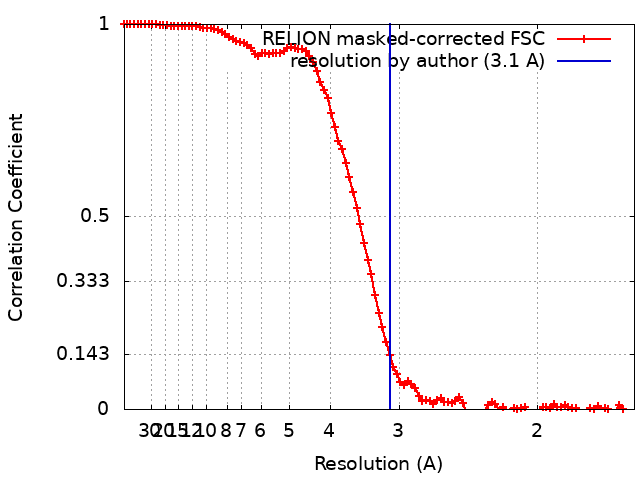
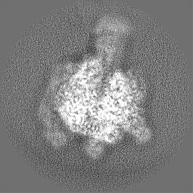
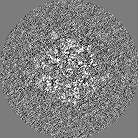
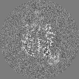
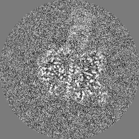
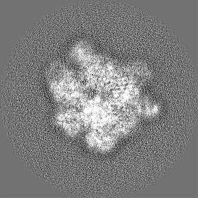
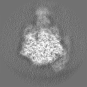
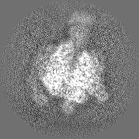
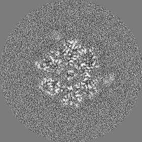
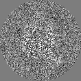
single particle reconstruction / cryo EM / Resolution: 3.1 Å
Ministry of Education, Culture, Sports, Science and Technology (Japan)
JPMXP09A21OS0008
Japan
Japan Agency for Medical Research and Development (AMED)
JP17am0101001
Japan
Citation
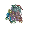
Journal: J Biol Chem / Year: 2023 Title: Cryo-EM analysis of V/A-ATPase intermediates reveals the transition of the ground-state structure to steady-state structures by sequential ATP binding. Authors: Atsuko Nakanishi / Jun-Ichi Kishikawa / Kaoru Mitsuoka / Ken Yokoyama / Abstract: Vacuolar/archaeal-type ATPase (V/A-ATPase) is a rotary ATPase that shares a common rotary catalytic mechanism with FF ATP synthase. Structural images of V/A-ATPase obtained by single-particle cryo- ...Vacuolar/archaeal-type ATPase (V/A-ATPase) is a rotary ATPase that shares a common rotary catalytic mechanism with FF ATP synthase. Structural images of V/A-ATPase obtained by single-particle cryo-electron microscopy during ATP hydrolysis identified several intermediates, revealing the rotary mechanism under steady-state conditions. However, further characterization is needed to understand the transition from the ground state to the steady state. Here, we identified the cryo-electron microscopy structures of V/A-ATPase corresponding to short-lived initial intermediates during the activation of the ground state structure by time-resolving snapshot analysis. These intermediate structures provide insights into how the ground-state structure changes to the active, steady state through the sequential binding of ATP to its three catalytic sites. All the intermediate structures of V/A-ATPase adopt the same asymmetric structure, whereas the three catalytic dimers adopt different conformations. This is significantly different from the initial activation process of FF, where the overall structure of the F domain changes during the transition from a pseudo-symmetric to a canonical asymmetric structure (PNAS NEXUS, pgac116, 2022). In conclusion, our findings provide dynamical information that will enhance the future prospects for studying the initial activation processes of the enzymes, which have unknown intermediate structures in their functional pathway.
Entire : 1 sulfate and 1 ATP bound V1EG of V/A-ATPase from Thermus thermophilus
Entire
Name: 1 sulfate and 1 ATP bound V1EG of V/A-ATPase from Thermus thermophilus
Components
Complex: 1 sulfate and 1 ATP bound V1EG of V/A-ATPase from Thermus thermophilus
Protein or peptide: V-type ATP synthase alpha chain
Protein or peptide: V-type ATP synthase beta chain
Protein or peptide: V-type ATP synthase subunit D
Protein or peptide: V-type ATP synthase subunit F
Protein or peptide: V-type ATP synthase, subunit (VAPC-THERM)
Protein or peptide: V-type ATP synthase subunit E
Ligand: SULFATE ION
Ligand: ADENOSINE-5'-TRIPHOSPHATE
Ligand: MAGNESIUM ION
+
Supramolecule #1: 1 sulfate and 1 ATP bound V1EG of V/A-ATPase from Thermus thermophilus
Supramolecule
Name: 1 sulfate and 1 ATP bound V1EG of V/A-ATPase from Thermus thermophilus type: complex / ID: 1 / Parent: 0 / Macromolecule list: #1-#6
Source (natural)
Organism: Thermus thermophilus HB8 (bacteria)
Molecular weight
Theoretical: 600 KDa
+
Macromolecule #1: V-type ATP synthase alpha chain
Macromolecule
Name: V-type ATP synthase alpha chain / type: protein_or_peptide / ID: 1 Details: Authors state that the bacterium they used has two mutations in its genome (S232A and T235S) and they obtained the EM sample from Natural source. Number of copies: 3 / Enantiomer: LEVO / EC number: H+-transporting two-sector ATPase
In the structure databanks used in Yorodumi, some data are registered as the other names, "COVID-19 virus" and "2019-nCoV". Here are the details of the virus and the list of structure data.
Jan 31, 2019. EMDB accession codes are about to change! (news from PDBe EMDB page)
EMDB accession codes are about to change! (news from PDBe EMDB page)
The allocation of 4 digits for EMDB accession codes will soon come to an end. Whilst these codes will remain in use, new EMDB accession codes will include an additional digit and will expand incrementally as the available range of codes is exhausted. The current 4-digit format prefixed with “EMD-” (i.e. EMD-XXXX) will advance to a 5-digit format (i.e. EMD-XXXXX), and so on. It is currently estimated that the 4-digit codes will be depleted around Spring 2019, at which point the 5-digit format will come into force.
The EM Navigator/Yorodumi systems omit the EMD- prefix.
Related info.:Q: What is EMD? / ID/Accession-code notation in Yorodumi/EM Navigator
Yorodumi is a browser for structure data from EMDB, PDB, SASBDB, etc.
This page is also the successor to EM Navigator detail page, and also detail information page/front-end page for Omokage search.
The word "yorodu" (or yorozu) is an old Japanese word meaning "ten thousand". "mi" (miru) is to see.
Related info.:EMDB / PDB / SASBDB / Comparison of 3 databanks / Yorodumi Search / Aug 31, 2016. New EM Navigator & Yorodumi / Yorodumi Papers / Jmol/JSmol / Function and homology information / Changes in new EM Navigator and Yorodumi
 Movie
Movie Controller

 Yorodumi
 Open data
 Basic information
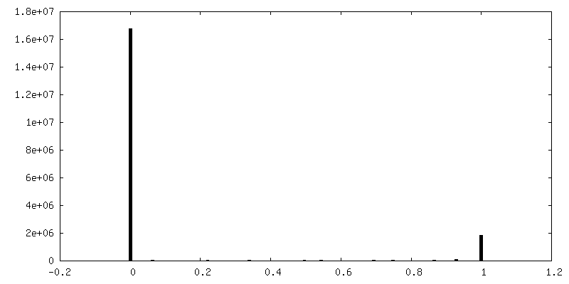
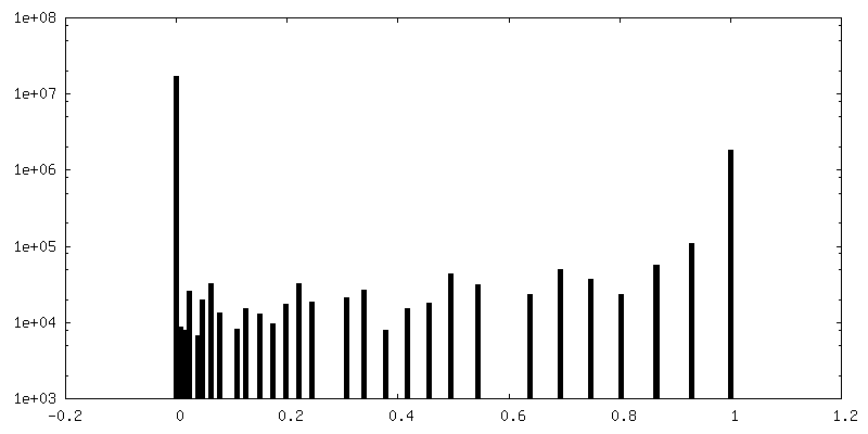
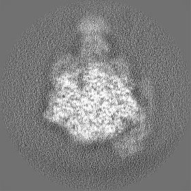
 Map data
 Sample
 Keywords
 Function and homology information
 Thermus thermophilus HB8 (bacteria)
 Authors
 Japan, 5 items
  Citation
 Structure visualization
 Downloads & links
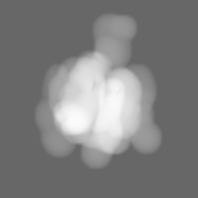
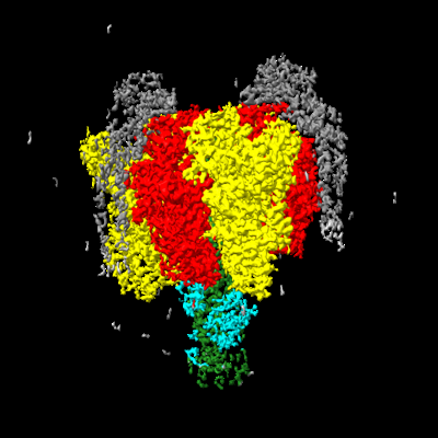 emd_34366.png
 http://ftp.pdbj.org/pub/emdb/structures/EMD-34366


 Z (Sec.)
 Y (Row.)
 X (Col.)


 Sample components

 Processing
 Electron microscopy
 FIELD EMISSION GUN