SOS response / RecA / UmuD / Filament / DNA repair / Helical Reconstruction / DNA BINDING PROTEIN-DNA COMPLEX
Function / homology
Function and homology information
repressor LexA / SOS response / ATP-dependent DNA damage sensor activity / Hydrolases; Acting on peptide bonds (peptidases); Serine endopeptidases / single-stranded DNA binding / DNA recombination / damaged DNA binding / DNA-directed DNA polymerase / DNA-directed DNA polymerase activity / hydrolase activity ...repressor LexA / SOS response / ATP-dependent DNA damage sensor activity / Hydrolases; Acting on peptide bonds (peptidases); Serine endopeptidases / single-stranded DNA binding / DNA recombination / damaged DNA binding / DNA-directed DNA polymerase / DNA-directed DNA polymerase activity / hydrolase activity / DNA repair / regulation of DNA-templated transcription / ATP hydrolysis activity / DNA binding / ATP binding / cytosol Similarity search - Function
Peptidase S24, LexA-like / LexA-like / Peptidase S24/S26A/S26B/S26C / Peptidase S24-like / LexA/Signal peptidase-like superfamily / : / : / RecA C-terminal domain / DNA recombination/repair protein RecA, conserved site / DNA recombination and repair protein RecA, C-terminal ...Peptidase S24, LexA-like / LexA-like / Peptidase S24/S26A/S26B/S26C / Peptidase S24-like / LexA/Signal peptidase-like superfamily / : / : / RecA C-terminal domain / DNA recombination/repair protein RecA, conserved site / DNA recombination and repair protein RecA, C-terminal / recA signature. / DNA recombination and repair protein RecA / recA bacterial DNA recombination protein / DNA recombination and repair protein RecA, monomer-monomer interface / RecA family profile 2. / DNA recombination and repair protein RecA-like, ATP-binding domain / RecA family profile 1. / ATPases associated with a variety of cellular activities / AAA+ ATPase domain / P-loop containing nucleoside triphosphate hydrolase Similarity search - Domain/homology
National Natural Science Foundation of China (NSFC)
31970040
China
Citation
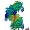
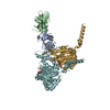
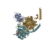
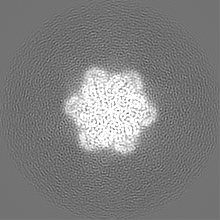
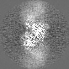
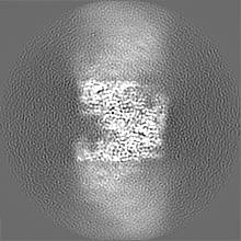
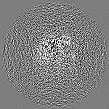
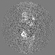
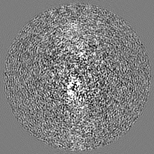
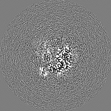
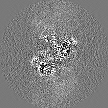
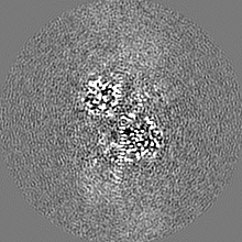
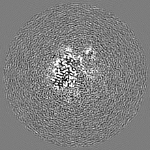
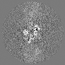
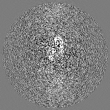
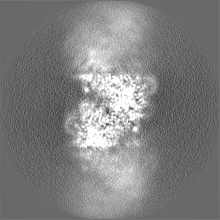
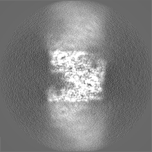
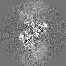
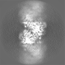
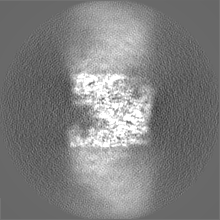
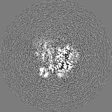
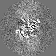
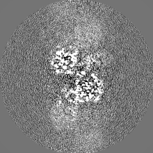
Journal: Proc Natl Acad Sci U S A / Year: 2023 Title: Structural basis for regulation of SOS response in bacteria. Authors: Bo Gao / Liang Liang / Lu Su / Aijia Wen / Chun Zhou / Yu Feng / Abstract: In response to DNA damage, bacterial RecA protein forms filaments with the assistance of DinI protein. The RecA filaments stimulate the autocleavage of LexA, the repressor of more than 50 SOS genes, ...In response to DNA damage, bacterial RecA protein forms filaments with the assistance of DinI protein. The RecA filaments stimulate the autocleavage of LexA, the repressor of more than 50 SOS genes, and activate the SOS response. During the late phase of SOS response, the RecA filaments stimulate the autocleavage of UmuD and λ repressor CI, leading to mutagenic repair and lytic cycle, respectively. Here, we determined the cryo-electron microscopy structures of RecA filaments in complex with DinI, LexA, UmuD, and λCI by helical reconstruction. The structures reveal that LexA and UmuD dimers bind in the filament groove and cleave in an intramolecular and an intermolecular manner, respectively, while λCI binds deeply in the filament groove as a monomer. Despite their distinct folds and oligomeric states, all RecA filament binders recognize the same conserved protein features in the filament groove. The SOS response in bacteria can lead to mutagenesis and antimicrobial resistance, and our study paves the way for rational drug design targeting the bacterial SOS response.
In the structure databanks used in Yorodumi, some data are registered as the other names, "COVID-19 virus" and "2019-nCoV". Here are the details of the virus and the list of structure data.
Jan 31, 2019. EMDB accession codes are about to change! (news from PDBe EMDB page)
EMDB accession codes are about to change! (news from PDBe EMDB page)
The allocation of 4 digits for EMDB accession codes will soon come to an end. Whilst these codes will remain in use, new EMDB accession codes will include an additional digit and will expand incrementally as the available range of codes is exhausted. The current 4-digit format prefixed with “EMD-” (i.e. EMD-XXXX) will advance to a 5-digit format (i.e. EMD-XXXXX), and so on. It is currently estimated that the 4-digit codes will be depleted around Spring 2019, at which point the 5-digit format will come into force.
The EM Navigator/Yorodumi systems omit the EMD- prefix.
Related info.:Q: What is EMD? / ID/Accession-code notation in Yorodumi/EM Navigator
Yorodumi is a browser for structure data from EMDB, PDB, SASBDB, etc.
This page is also the successor to EM Navigator detail page, and also detail information page/front-end page for Omokage search.
The word "yorodu" (or yorozu) is an old Japanese word meaning "ten thousand". "mi" (miru) is to see.
Related info.:EMDB / PDB / SASBDB / Comparison of 3 databanks / Yorodumi Search / Aug 31, 2016. New EM Navigator & Yorodumi / Yorodumi Papers / Jmol/JSmol / Function and homology information / Changes in new EM Navigator and Yorodumi
 Movie
Movie Controller


 Open data
 Basic information
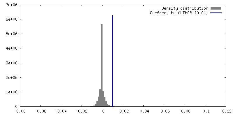
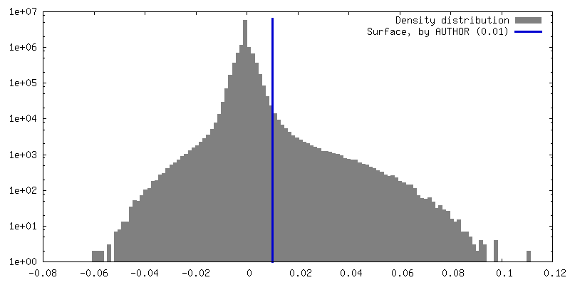
 Map data
 Sample
 Keywords
 Function and homology information
 Authors
 China, 1 items
  Citation
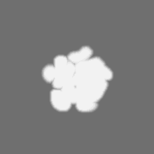
 Structure visualization
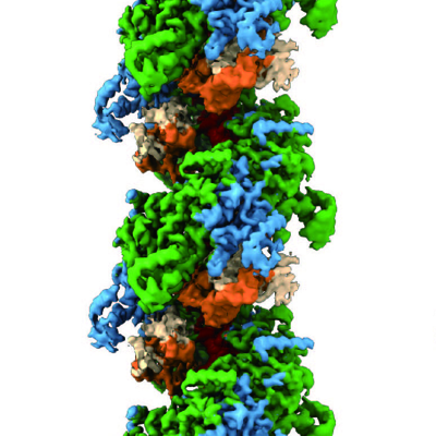
 Downloads & links
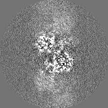
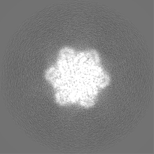
 emd_34153.png
 http://ftp.pdbj.org/pub/emdb/structures/EMD-34153


 Z (Sec.)
 Y (Row.)
 X (Col.)


 Sample components
 Processing
 Electron microscopy
 FIELD EMISSION GUN