Movie
Movie Controller

[English] 日本語
 Yorodumi
- EMDB-33940: Structure of recombinant RyR2 (Ca2+ dataset, class 3, open state) -
+ Open data

- Basic information
| Entry |  | |||||||||||||||||||||
|---|---|---|---|---|---|---|---|---|---|---|---|---|---|---|---|---|---|---|---|---|---|---|
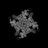
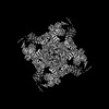
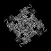
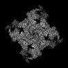
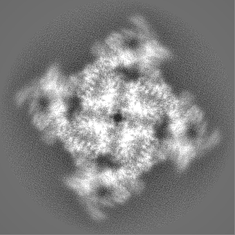
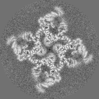
| Title | Structure of recombinant RyR2 (Ca2+ dataset, class 3, open state) | |||||||||||||||||||||
 Map data Map data | Structure of recombinant RyR2 (Ca2 dataset, class 3, open state) | |||||||||||||||||||||
 Sample Sample |
| |||||||||||||||||||||
| Function / homology |  Function and homology information Function and homology informationmanganese ion transmembrane transport / establishment of protein localization to endoplasmic reticulum / type B pancreatic cell apoptotic process / Purkinje myocyte to ventricular cardiac muscle cell signaling / regulation of atrial cardiac muscle cell action potential / left ventricular cardiac muscle tissue morphogenesis / suramin binding / regulation of AV node cell action potential / regulation of SA node cell action potential / sarcoplasmic reticulum calcium ion transport ...manganese ion transmembrane transport / establishment of protein localization to endoplasmic reticulum / type B pancreatic cell apoptotic process / Purkinje myocyte to ventricular cardiac muscle cell signaling / regulation of atrial cardiac muscle cell action potential / left ventricular cardiac muscle tissue morphogenesis / suramin binding / regulation of AV node cell action potential / regulation of SA node cell action potential / sarcoplasmic reticulum calcium ion transport / A band / calcium-induced calcium release activity / Stimuli-sensing channels / regulation of ventricular cardiac muscle cell action potential / : / ventricular cardiac muscle cell action potential / embryonic heart tube morphogenesis / negative regulation of calcium-mediated signaling / Ion homeostasis / cardiac muscle hypertrophy / negative regulation of insulin secretion involved in cellular response to glucose stimulus / calcium ion transport into cytosol / neuronal action potential propagation / insulin secretion involved in cellular response to glucose stimulus / negative regulation of release of sequestered calcium ion into cytosol / ryanodine-sensitive calcium-release channel activity / response to caffeine / release of sequestered calcium ion into cytosol by sarcoplasmic reticulum / response to redox state / regulation of cardiac muscle contraction by calcium ion signaling / negative regulation of heart rate / cellular response to caffeine / extrinsic component of cytoplasmic side of plasma membrane / 'de novo' protein folding / calcium ion transmembrane import into cytosol / FK506 binding / response to muscle activity / negative regulation of cytosolic calcium ion concentration / protein kinase A regulatory subunit binding / protein kinase A catalytic subunit binding / positive regulation of the force of heart contraction / intracellularly gated calcium channel activity / smooth endoplasmic reticulum / smooth muscle contraction / response to magnesium ion / T cell proliferation / detection of calcium ion / regulation of cardiac muscle contraction / positive regulation of heart rate / regulation of cytosolic calcium ion concentration / calcium channel inhibitor activity / striated muscle contraction / Ion homeostasis / regulation of release of sequestered calcium ion into cytosol by sarcoplasmic reticulum / cardiac muscle contraction / response to muscle stretch / release of sequestered calcium ion into cytosol / regulation of cardiac muscle contraction by regulation of the release of sequestered calcium ion / sarcoplasmic reticulum membrane / cellular response to epinephrine stimulus / calcium channel complex / regulation of heart rate / sarcomere / sarcoplasmic reticulum / peptidylprolyl isomerase / peptidyl-prolyl cis-trans isomerase activity / calcium channel regulator activity / calcium-mediated signaling / protein maturation / response to calcium ion / sarcolemma / Stimuli-sensing channels / calcium ion transmembrane transport / calcium channel activity / Z disc / intracellular calcium ion homeostasis / calcium ion transport / nuclear envelope / protein refolding / positive regulation of cytosolic calcium ion concentration / scaffold protein binding / monoatomic ion transmembrane transport / transmembrane transporter binding / response to hypoxia / calmodulin binding / signaling receptor binding / calcium ion binding / protein kinase binding / enzyme binding / protein-containing complex / membrane / identical protein binding / cytoplasm Similarity search - Function | |||||||||||||||||||||
| Biological species |   Homo sapiens (human) Homo sapiens (human) | |||||||||||||||||||||
| Method | single particle reconstruction / cryo EM / Resolution: 3.72 Å | |||||||||||||||||||||
 Authors Authors | Kobayashi T / Tsutsumi A / Kurebayashi N / Kodama M / Kikkawa M / Murayama T / Ogawa H | |||||||||||||||||||||
| Funding support |  Japan, 6 items Japan, 6 items
| |||||||||||||||||||||
 Citation Citation | Journal: Nat Commun / Year: 2022 Title: Molecular basis for gating of cardiac ryanodine receptor explains the mechanisms for gain- and loss-of function mutations. Authors: Takuya Kobayashi / Akihisa Tsutsumi / Nagomi Kurebayashi / Kei Saito / Masami Kodama / Takashi Sakurai / Masahide Kikkawa / Takashi Murayama / Haruo Ogawa / Abstract: Cardiac ryanodine receptor (RyR2) is a large Ca release channel in the sarcoplasmic reticulum and indispensable for excitation-contraction coupling in the heart. RyR2 is activated by Ca and RyR2 ...Cardiac ryanodine receptor (RyR2) is a large Ca release channel in the sarcoplasmic reticulum and indispensable for excitation-contraction coupling in the heart. RyR2 is activated by Ca and RyR2 mutations are implicated in severe arrhythmogenic diseases. Yet, the structural basis underlying channel opening and how mutations affect the channel remains unknown. Here, we address the gating mechanism of RyR2 by combining high-resolution structures determined by cryo-electron microscopy with quantitative functional analysis of channels carrying various mutations in specific residues. We demonstrated two fundamental mechanisms for channel gating: interactions close to the channel pore stabilize the channel to prevent hyperactivity and a series of interactions in the surrounding regions is necessary for channel opening upon Ca binding. Mutations at the residues involved in the former and the latter mechanisms cause gain-of-function and loss-of-function, respectively. Our results reveal gating mechanisms of the RyR2 channel and alterations by pathogenic mutations at the atomic level. | |||||||||||||||||||||
| History |
|
- Structure visualization
| Supplemental images |
|---|

- Downloads & links
-EMDB archive
| Map data | emd_33940.map.gz | 138.9 MB | EMDB map data format | |
|---|---|---|---|---|
| Header (meta data) | emd-33940-v30.xmlemd-33940.xml | 15.7 KB 15.7 KB | Display Display | EMDB header |
| Images | 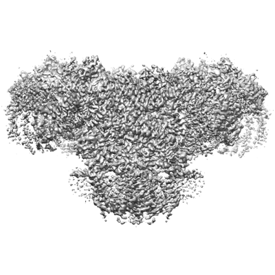 emd_33940.png emd_33940.png | 125.5 KB | ||
| Others | emd_33940_half_map_1.map.gzemd_33940_half_map_2.map.gz | 113.4 MB 113.3 MB | ||
| Archive directory |  http://ftp.pdbj.org/pub/emdb/structures/EMD-33940ftp://ftp.pdbj.org/pub/emdb/structures/EMD-33940 http://ftp.pdbj.org/pub/emdb/structures/EMD-33940ftp://ftp.pdbj.org/pub/emdb/structures/EMD-33940 | HTTPS FTP |
-Related structure data
| Related structure data |  7vmqM  7vmlC  7vmmC  7vmnC  7vmoC  7vmpC  7vmrC M: atomic model generated by this map C: citing same article ( |
|---|---|
| Similar structure data |

-Links
| EMDB pages | EMDB (EBI/PDBe) / EMDataResource |
|---|---|
| Related items in Molecule of the Month |


-Map
| File | Download / File: emd_33940.map.gz / Format: CCP4 / Size: 149.9 MB / Type: IMAGE STORED AS FLOATING POINT NUMBER (4 BYTES) | ||||||||||||||||||||||||||||||||||||
|---|---|---|---|---|---|---|---|---|---|---|---|---|---|---|---|---|---|---|---|---|---|---|---|---|---|---|---|---|---|---|---|---|---|---|---|---|---|
| Annotation | Structure of recombinant RyR2 (Ca2 dataset, class 3, open state) | ||||||||||||||||||||||||||||||||||||
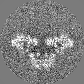
| Projections & slices | Image control
Images are generated by Spider. | ||||||||||||||||||||||||||||||||||||
| Voxel size | X=Y=Z: 1.239 Å | ||||||||||||||||||||||||||||||||||||
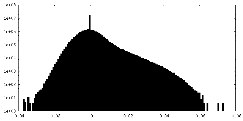
| Density |
| ||||||||||||||||||||||||||||||||||||
| Symmetry | Space group: 1 | ||||||||||||||||||||||||||||||||||||
| Details | EMDB XML:
|
 Z (Sec.)
 Y (Row.)
 X (Col.)


-Supplemental data
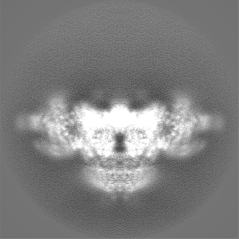
-Half map: Structure of recombinant RyR2 (Ca2 dataset, class 3, open state)
| File | emd_33940_half_map_1.map | ||||||||||||
|---|---|---|---|---|---|---|---|---|---|---|---|---|---|
| Annotation | Structure of recombinant RyR2 (Ca2 dataset, class 3, open state) | ||||||||||||
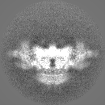
| Projections & Slices |
| ||||||||||||
| Density Histograms |
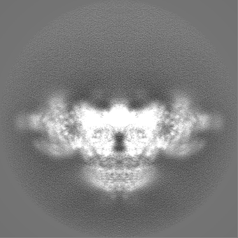


-Half map: Structure of recombinant RyR2 (Ca2 dataset, class 3, open state)
| File | emd_33940_half_map_2.map | ||||||||||||
|---|---|---|---|---|---|---|---|---|---|---|---|---|---|
| Annotation | Structure of recombinant RyR2 (Ca2 dataset, class 3, open state) | ||||||||||||
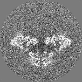
| Projections & Slices |
| ||||||||||||
| Density Histograms |


- Sample components
-Entire : Recombinant RyR2 in the presence of EGTA
| Entire | Name: Recombinant RyR2 in the presence of EGTA |
|---|---|
| Components |
|
-Supramolecule #1: Recombinant RyR2 in the presence of EGTA
| Supramolecule | Name: Recombinant RyR2 in the presence of EGTA / type: complex / Chimera: Yes / ID: 1 / Parent: 0 / Macromolecule list: #1-#2 / Details: in complex with FKBP12.6 |
|---|---|
| Source (natural) | Organism: |
| Recombinant expression | Organism: Homo sapiens (human) |
-Supramolecule #2: Ryanodine receptor 2
| Supramolecule | Name: Ryanodine receptor 2 / type: complex / ID: 2 / Parent: 1 / Macromolecule list: #1 |
|---|---|
| Source (natural) | Organism: Homo sapiens (human) |
| Recombinant expression | Organism:  |
-Supramolecule #3: FKBP1B
| Supramolecule | Name: FKBP1B / type: complex / ID: 3 / Parent: 1 / Macromolecule list: #2 |
|---|
-Experimental details
-Structure determination
| Method | cryo EM |
|---|---|
 Processing Processing | single particle reconstruction |
| Aggregation state | particle |
-Sample preparation
| Buffer | pH: 7.4 |
|---|---|
| Sugar embedding | Material: buffer |
| Vitrification | Cryogen name: ETHANE |
- Electron microscopy
| Microscope | FEI TITAN KRIOS |
|---|---|
| Image recording | Film or detector model: GATAN K3 (6k x 4k) / Average electron dose: 60.0 e/Å2 |
| Electron beam | Acceleration voltage: 300 kV / Electron source:  FIELD EMISSION GUN FIELD EMISSION GUN |
| Electron optics | Illumination mode: FLOOD BEAM / Imaging mode: BRIGHT FIELD / Nominal defocus max: 2.0 µm / Nominal defocus min: 0.5 µm |
| Experimental equipment |  Model: Titan Krios / Image courtesy: FEI Company |
-Image processing
| Startup model | Type of model: PDB ENTRY PDB model - PDB ID: |
|---|---|
| Final reconstruction | Resolution.type: BY AUTHOR / Resolution: 3.72 Å / Resolution method: FSC 0.143 CUT-OFF / Number images used: 40665 |
| Initial angle assignment | Type: MAXIMUM LIKELIHOOD |
| Final angle assignment | Type: MAXIMUM LIKELIHOOD |