[English] 日本語
Yorodumi
- EMDB-32352: Structural of the filamentous Escherichia coli glutamine synthetase -

+
Open data


ID or keywords:

Loading...

-
Basic information

Entry
Database: EMDB / ID: EMD-32352
TitleStructural of the filamentous Escherichia coli glutamine synthetase
Map dataSPA map of Ecoli GS filament, calculated using cryoSPARC.
Sample
  • Complex: Filamentous form of Ecoli glutamine synthetase
    • Protein or peptide: Glutamine synthetase
  • Ligand: NICKEL (II) ION
Function / homology
Function and homology information


ammonia assimilation cycle / nitrogen utilization / glutamine synthetase / glutamine biosynthetic process / glutamine synthetase activity / response to radiation / ATP binding / membrane / identical protein binding / metal ion binding ...ammonia assimilation cycle / nitrogen utilization / glutamine synthetase / glutamine biosynthetic process / glutamine synthetase activity / response to radiation / ATP binding / membrane / identical protein binding / metal ion binding / cytosol / cytoplasm
Similarity search - Function
Glutamine synthetase class-I, adenylation site / Glutamine synthetase class-I adenylation site. / Glutamine synthetase type I / Glutamine synthetase (GS) beta-grasp domain profile. / Glutamine synthetase (GS) catalytic domain profile. / Glutamine synthetase, N-terminal conserved site / Glutamine synthetase signature 1. / Glutamine synthetase, beta-Grasp domain / Glutamine synthetase, glycine-rich site / Glutamine synthetase putative ATP-binding region signature. ...Glutamine synthetase class-I, adenylation site / Glutamine synthetase class-I adenylation site. / Glutamine synthetase type I / Glutamine synthetase (GS) beta-grasp domain profile. / Glutamine synthetase (GS) catalytic domain profile. / Glutamine synthetase, N-terminal conserved site / Glutamine synthetase signature 1. / Glutamine synthetase, beta-Grasp domain / Glutamine synthetase, glycine-rich site / Glutamine synthetase putative ATP-binding region signature. / Glutamine synthetase, N-terminal domain / Glutamine synthetase, N-terminal domain superfamily / Glutamine synthetase, catalytic domain / Glutamine synthetase, catalytic domain / Glutamine synthetase, catalytic domain / Glutamine synthetase/guanido kinase, catalytic domain
Similarity search - Domain/homology
Glutamine synthetase
Similarity search - Component
Biological speciesEscherichia coli BL21(DE3) (bacteria)
Methodsingle particle reconstruction / cryo EM / Resolution: 2.94 Å
AuthorsHuang P-C / Chen S-K / Wu K-P
Funding support Taiwan, 2 items
OrganizationGrant numberCountry
Ministry of Science and Technology (MoST, Taiwan) Taiwan
Academia Sinica (Taiwan) Taiwan
Citation
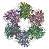
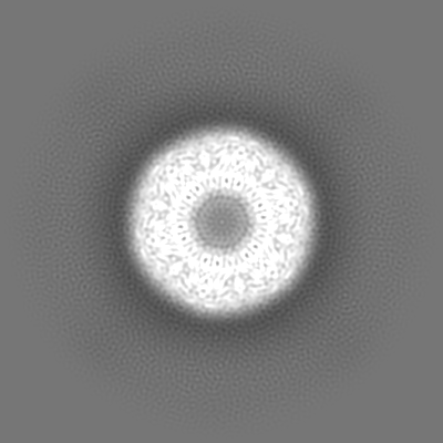
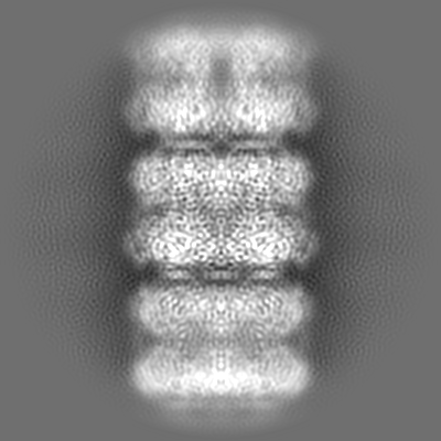
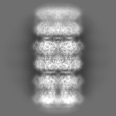
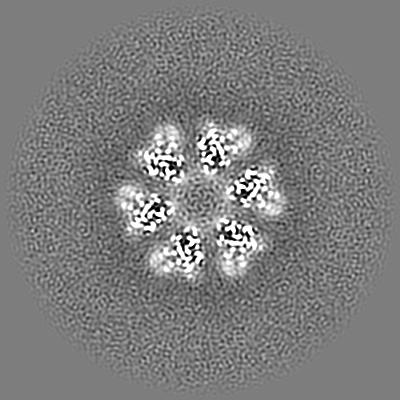
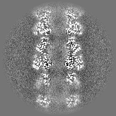
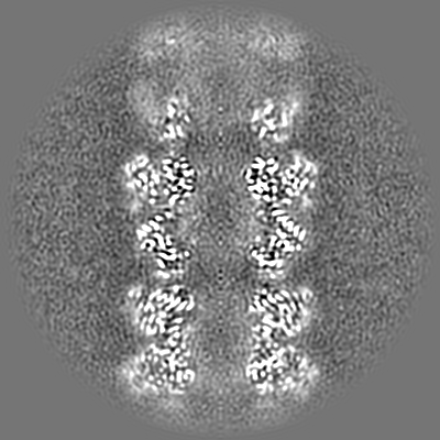
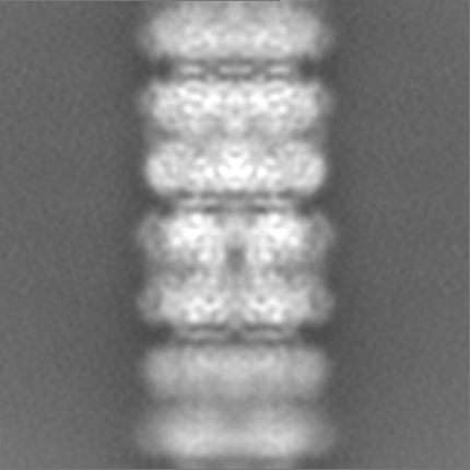
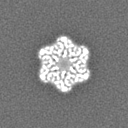
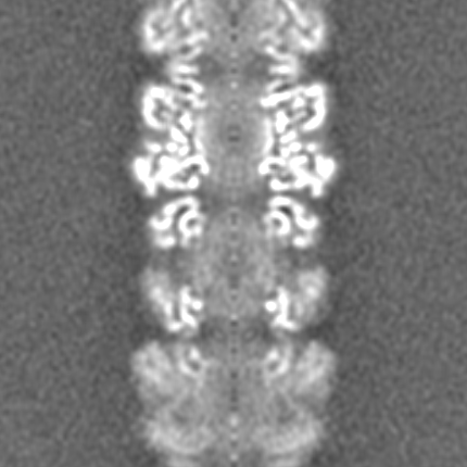
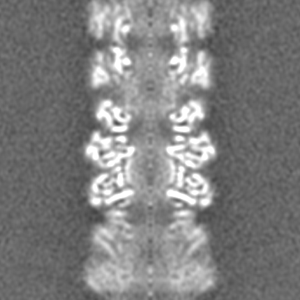
Journal: Protein Sci / Year: 2022
Title: Structural basis for the helical filament formation of Escherichia coli glutamine synthetase.
Authors: Pei-Chi Huang / Shao-Kang Chen / Wei-Hung Chiang / Meng-Ru Ho / Kuen-Phon Wu /
Abstract: Escherichia coli glutamine synthetase (EcGS) spontaneously forms a dodecamer that catalytically converts glutamate to glutamine. EcGS stacks with other dodecamers to create a filament-like polymer ...Escherichia coli glutamine synthetase (EcGS) spontaneously forms a dodecamer that catalytically converts glutamate to glutamine. EcGS stacks with other dodecamers to create a filament-like polymer visible under transmission electron microscopy. Filamentous EcGS is induced by environmental metal ions. We used cryo-electron microscopy (cryo-EM) to decipher the structure of metal ion (nickel)-induced EcGS helical filament at a sub-3Å resolution. EcGS filament formation involves stacking of native dodecamers by chelating nickel ions to residues His5 and His13 in the first N-terminal helix (H1). His5 and His13 from paired parallel H1 helices provide salt bridges and hydrogen bonds to tightly stack two dodecamers. One subunit of the EcGS filament hosts two nickel ions, whereas the dodecameric interface and the ATP/Mg-binding site both host a nickel ion each. We reveal that upon adding glutamate or ATP for catalytic reactions, nickel-induced EcGS filament reverts to individual dodecamers. Such tunable filament formation is often associated with stress responses. Our results provide detailed structural information on the mechanism underlying reversible and tunable EcGS filament formation.
History
DepositionDec 7, 2021-
Header (metadata) releaseApr 6, 2022-
Map releaseApr 6, 2022-
UpdateJun 15, 2022-
Current statusJun 15, 2022Processing site: PDBj / Status: Released

-
Structure visualization
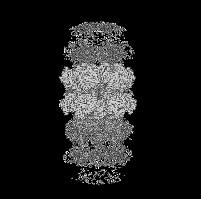
Supplemental images

Downloads & links

-
Map

FileDownload / File: emd_32352.map.gz / Format: CCP4 / Size: 303.3 MB / Type: IMAGE STORED AS FLOATING POINT NUMBER (4 BYTES)
AnnotationSPA map of Ecoli GS filament, calculated using cryoSPARC.
Voxel sizeX=Y=Z: 0.822 Å
Density
Contour LevelBy AUTHOR: 0.4
Minimum - Maximum-2.135863 - 3.2281482
Average (Standard dev.)0.020110242 (±0.09557043)
SymmetrySpace group: 1
Details

EMDB XML:

Map geometry
Axis orderXYZ
Origin000
Dimensions430430430
Spacing430430430
CellA=B=C: 353.46002 Å
α=β=γ: 90.0 °

-
Supplemental data

-
Additional map: Helical reconstruction map of Ecoli GS filament, calculated...

Fileemd_32352_additional_1.map
AnnotationHelical reconstruction map of Ecoli GS filament, calculated using cryoSPARC.
Projections & Slices
AxesZYX

Projections

Slices (1/2)
Density Histograms

-
Additional map: additional map for the 24mer (12mer x2) assembly....

Fileemd_32352_additional_2.map
Annotationadditional map for the 24mer (12mer x2) assembly. It was calculated to 5.5 angstrom.
Projections & Slices
AxesZYX

Projections

Slices (1/2)
Density Histograms

-
Sample components

-
Entire : Filamentous form of Ecoli glutamine synthetase

EntireName: Filamentous form of Ecoli glutamine synthetase
Components
  • Complex: Filamentous form of Ecoli glutamine synthetase
    • Protein or peptide: Glutamine synthetase
  • Ligand: NICKEL (II) ION

-
Supramolecule #1: Filamentous form of Ecoli glutamine synthetase

SupramoleculeName: Filamentous form of Ecoli glutamine synthetase / type: complex / Chimera: Yes / ID: 1 / Parent: 0 / Macromolecule list: #1
Source (natural)Organism: Escherichia coli BL21(DE3) (bacteria)
Recombinant expressionOrganism: Escherichia coli BL21(DE3) (bacteria)

-
Macromolecule #1: Glutamine synthetase

MacromoleculeName: Glutamine synthetase / type: protein_or_peptide / ID: 1 / Number of copies: 24 / Enantiomer: LEVO / EC number: glutamine synthetase
Source (natural)Organism: Escherichia coli BL21(DE3) (bacteria) / Strain: K12
Molecular weightTheoretical: 51.966609 KDa
Recombinant expressionOrganism: Escherichia coli BL21(DE3) (bacteria)
SequenceString: MSAEHVLTML NEHEVKFVDL RFTDTKGKEQ HVTIPAHQVN AEFFEEGKMF DGSSIGGWKG INESDMVLMP DASTAVIDPF FADSTLIIR CDILEPGTLQ GYDRDPRSIA KRAEDYLRST GIADTVLFGP EPEFFLFDDI RFGSSISGSH VAIDDIEGAW N SSTQYEGG ...String:
MSAEHVLTML NEHEVKFVDL RFTDTKGKEQ HVTIPAHQVN AEFFEEGKMF DGSSIGGWKG INESDMVLMP DASTAVIDPF FADSTLIIR CDILEPGTLQ GYDRDPRSIA KRAEDYLRST GIADTVLFGP EPEFFLFDDI RFGSSISGSH VAIDDIEGAW N SSTQYEGG NKGHRPAVKG GYFPVPPVDS AQDIRSEMCL VMEQMGLVVE AHHHEVATAG QNEVATRFNT MTKKADEIQI YK YVVHNVA HRFGKTATFM PKPMFGDNGS GMHCHMSLSK NGVNLFAGDK YAGLSEQALY YIGGVIKHAK AINALANPTT NSY KRLVPG YEAPVMLAYS ARNRSASIRI PVVSSPKARR IEVRFPDPAA NPYLCFAALL MAGLDGIKNK IHPGEAMDKN LYDL PPEEA KEIPQVAGSL EEALNELDLD REFLKAGGVF TDEAIDAYIA LRREEDDRVR MTPHPVEFEL YYSV

-
Macromolecule #2: NICKEL (II) ION

MacromoleculeName: NICKEL (II) ION / type: ligand / ID: 2 / Number of copies: 42 / Formula: NI
Molecular weightTheoretical: 58.693 Da
Chemical component information

ChemComp-NI:
NICKEL (II) ION / Nickel

-
Experimental details

-
Structure determination

Methodcryo EM
Processingsingle particle reconstruction
Aggregation stateparticle

-
Sample preparation

Concentration0.1 mg/mL
BufferpH: 7.4
GridModel: Quantifoil R2/1 / Material: COPPER / Mesh: 200
VitrificationCryogen name: ETHANE / Instrument: FEI VITROBOT MARK IV
DetailsTitan Krios

-
Electron microscopy

MicroscopeTFS KRIOS
Electron beamAcceleration voltage: 300 kV / Electron source: FIELD EMISSION GUN
Electron opticsIllumination mode: FLOOD BEAM / Imaging mode: BRIGHT FIELDBright-field microscopy / Nominal defocus max: 2.0 µm / Nominal defocus min: 1.0 µm
Image recordingFilm or detector model: GATAN K2 SUMMIT (4k x 4k) / Detector mode: COUNTING / Number grids imaged: 1 / Number real images: 1182 / Average electron dose: 59.6 e/Å2
Experimental equipment
Model: Titan Krios / Image courtesy: FEI Company

-
Image processing

CTF correctionSoftware: (Name: cryoSPARC (ver. 2.14), cryoSPARC (ver. 3.2))
Initial angle assignmentType: MAXIMUM LIKELIHOOD / Software - Name: cryoSPARC (ver. 3.2)
Final angle assignmentType: MAXIMUM LIKELIHOOD / Software - Name: cryoSPARC (ver. 3.2)
Final reconstructionApplied symmetry - Point group: C6 (6 fold cyclic) / Resolution.type: BY AUTHOR / Resolution: 2.94 Å / Resolution method: FSC 0.143 CUT-OFF / Software - Name: cryoSPARC (ver. 3.2) / Number images used: 117242

-
Atomic model buiding 1

Initial modelPDB ID:

Chain - Chain ID: A
RefinementSpace: REAL / Protocol: RIGID BODY FIT
Output model

PDB-7w85:
Structural of the filamentous Escherichia coli glutamine synthetase

+
About Yorodumi

-
News

-
Feb 9, 2022. New format data for meta-information of EMDB entries

New format data for meta-information of EMDB entries

  • Version 3 of the EMDB header file is now the official format.
  • The previous official version 1.9 will be removed from the archive.

Related info.:EMDB header

External links:wwPDB to switch to version 3 of the EMDB data model

-
Aug 12, 2020. Covid-19 info

Covid-19 info

URL: https://pdbj.org/emnavi/covid19.php

New page: Covid-19 featured information page in EM Navigator.

Related info.:Covid-19 info / Mar 5, 2020. Novel coronavirus structure data

+
Mar 5, 2020. Novel coronavirus structure data

Novel coronavirus structure data

Related info.:Yorodumi Speices / Aug 12, 2020. Covid-19 info

External links:COVID-19 featured content - PDBj / Molecule of the Month (242):Coronavirus Proteases

+
Jan 31, 2019. EMDB accession codes are about to change! (news from PDBe EMDB page)

EMDB accession codes are about to change! (news from PDBe EMDB page)

  • The allocation of 4 digits for EMDB accession codes will soon come to an end. Whilst these codes will remain in use, new EMDB accession codes will include an additional digit and will expand incrementally as the available range of codes is exhausted. The current 4-digit format prefixed with “EMD-” (i.e. EMD-XXXX) will advance to a 5-digit format (i.e. EMD-XXXXX), and so on. It is currently estimated that the 4-digit codes will be depleted around Spring 2019, at which point the 5-digit format will come into force.
  • The EM Navigator/Yorodumi systems omit the EMD- prefix.

Related info.:Q: What is EMD? / ID/Accession-code notation in Yorodumi/EM Navigator

External links:EMDB Accession Codes are Changing Soon! / Contact to PDBj

+
Jul 12, 2017. Major update of PDB

Major update of PDB

  • wwPDB released updated PDB data conforming to the new PDBx/mmCIF dictionary.
  • This is a major update changing the version number from 4 to 5, and with Remediation, in which all the entries are updated.
  • In this update, many items about electron microscopy experimental information are reorganized (e.g. em_software).
  • Now, EM Navigator and Yorodumi are based on the updated data.

External links:wwPDB Remediation / Enriched Model Files Conforming to OneDep Data Standards Now Available in the PDB FTP Archive

-
Yorodumi

Thousand views of thousand structures

  • Yorodumi is a browser for structure data from EMDB, PDB, SASBDB, etc.
  • This page is also the successor to EM Navigator detail page, and also detail information page/front-end page for Omokage search.
  • The word "yorodu" (or yorozu) is an old Japanese word meaning "ten thousand". "mi" (miru) is to see.

Related info.:EMDB / PDB / SASBDB / Comparison of 3 databanks / Yorodumi Search / Aug 31, 2016. New EM Navigator & Yorodumi / Yorodumi Papers / Jmol/JSmol / Function and homology information / Changes in new EM Navigator and Yorodumi

Read more